Download

1 / 22
220 likes | 392 Vues
Rehabilitation Faculty. Semnan University of Medical Sciences. Ankle Joint Kinesiology. Amir H. Bakhtiary PhD, PT Associate Professor. Physiotherapy Department Rehabilitation faculty Semnan University of Medical Sciences. Interphalangeal Joint. Synovial hing Joint

E N D
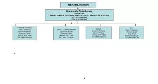
Rehabilitation Faculty Semnan University of Medical Sciences
Ankle Joint Kinesiology Amir H. Bakhtiary PhD, PT Associate Professor Physiotherapy Department Rehabilitation faculty Semnan University of Medical Sciences
Interphalangeal Joint • Synovial hing Joint • 1 degree of freedom (Fle/Ext) • 5 PIP Joint • 4 DIP • IP Joint function • Provide smooth WB transfer on the other foot • Provide stability by pressing the toes on the ground during standing and gait
Foot Arches • Provide by bone and ligament and Joint structure of foot • MTP and toes do NOT shear in the arches • But their position affect the curves • The foot arches is build by a twisted plan of foot in the TCN, Midtarsal and TMT Joint • The plan is in vertical position in calcaneous and • is in horizontal position in the metatarsus
Function of Foot Arches • Weight distribution • Force and Stress Absorbing • Force saving in its structure • Release saved energy at the end of stance phase This released energy used for walking Energy Saving System
Foot Arches • Longitudinal Arches • Medial (between calcaneous and first metatarsal) • More Higher and more flexibility • Lateral (between Calcaneous and lateral metatarsal) • More harder Less Higher and flexibility • Transverse Arch • From tarsus to the head of metatarsal • Higher in back • Lower in the front
Foot arches supporter factors • Ligaments • Plantar CalcaneonavicularLig (spring Lig) • Superior medial calcaneonavicular • InteroseosTalocalcaneoLig • Plantar aponeuroses • Cervical Lig • Long and Short plantar Lig • Muscles • PeroneusLongus • Flexor DigitrumLongus • Flexor HallusisLongus
The Role of Plantar Apponeurosis to Supportthe Longitudinal Arch
Plantar AponeourosesFunction • Act like a firm rope to hold the height of longitudinal arch • By stretching PA, the rear and front base of arch move toward each other and height will be increased • Decreased tension in PA • By WB the rear and front base of arch separate each other and height will be decreased and cause: • Increased tension in PA • Decreased Ext ROM in MTP joint
Effect Of Tibia Rotation on the height of arch foot in WB • Medial Rotation of Tibia cause • Pronation in Hindfoot • Decreased height of arch • Decreased Ext ROM in MTP joint • Lateral Rotation of Tibia cause • Supination in hindfoot • Increased height of arch • Increased Ext ROM in MTP joint These processes will be reversed
Foot WB Distribution • It depends on the • Shape of foot arches • Location of LOG • Talus received Body Weight • %50 in the bilateral standing • %100 in the unilateral standing • During standing, weight Transfer • %50 of weight to calcareous • %33 to Navicular • %17 to Cuboids • First Metatarsal Head distribute Weight 2 times more than other metatarsals
Foot WB Distribution • Pattern of weight distribution will differ during walking • In metatarsal break, Increase WB on the metatarsals • More WB on the II, III and IV metatarsals • Less WB on the I and V metatarsals • In Heel Contact, calcaneous receive 85-%100 • Fat pad help to reduce shocked from WB • During running, Received weight on the foot increased by the %250 of WB
Muscles which act on the foot * These muscle produce %50 of PF force • Extrinsic muscles • Plantar flexors • Gastrocnemious • Soleus • Plantaris* • Tibialis posterior* • Flexor Hallucis Longus* • Flexor Digitrum Longus* • Peroneus Longus* • Peroneus Brevis* • Dorsi flexors • Intrinsic muscles These muscle produce PF force & Supination
Muscles which act on the foot • سه سر ساق (گاستروکنمیوس و سولئوس) • از طریق آشیل به کالکانئوس متصل شده • دارای MA خوبی از مرکز چرخش مچ پا است • دارای اثر Sup است چون از داخل محور مچ پا می گذرد • موجب قفل شدن مفاصل TCN و Midtarsalپا هنگام PF • تبدیل پا به یک اهرم محکم • انقباض این عضلات پاشنه را از زمین بلند کرده • بلندتر شدن ارتفاع قوسهای کف پا در WB
Activity of the Triceps Surae • Great MA • Cause Sup in Hindfoot • In WB cause lock in TCN and Midtarsal • Make foot as a hard lever • Raise heel • Increase height of arches