Download
1 / 64
640 likes | 731 Vues
Chapter 9. List the organs and divisions of the nervous system and describe the generalized functions of the system as a whole. Identify the major types of cell in the nervous system and discuss the functions of each,

E N D
List the organs and divisions of the nervous system and describe the generalized functions of the system as a whole. • Identify the major types of cell in the nervous system and discuss the functions of each, • Identify the anatomical and functional components of a three-neuron reflex arc. Compare and contrast the propagation of a never impulse. • Identify the major anatomical components of the brain and spinal cord and briefly comment on the function of each. • Identify and discuss the coverings and fluid spaces of the brain and spinal cord. • Compare and contrast spinal and cranial nerves. • Discuss the structure and function of the two divisions of the autonomic nervous system. • Describe major nervous system disorders.
ORGANS AND DIVISIONS OF THE NERVOUS SYSTEM • CELLS OF THE NERVOUS SYSTEM • Neurons • Glia • Disorders of Nervous Tissue • NERVES • REFLEX ARCS • NERVE IMPULSES • THE SYNAPSE • CENTRAL NERVOUS SYSTEM • Divisions of the Brain • Brain Disorders • Spinal Cord • Coverings and Fluid Spaces of the Brain and Spinal Cord • PERIPHERAL NERVOUS SYSTEM • Cranial Nerves • Spinal nerves • Peripheral Nerve Disorders • AUTONOMIC NERVOUS SYSTEM • Functional Anatomy • Autonomic Conduction Paths • Sympathetic Nervous System • Parasympathetic Nervous System • Autonomic Neurotransmitters • Autonomic Nervous System as a Whole • Disorders of the Autonomic Nervous System
Organs and Divisions of the Nervous System (Figure 9-1) • Central nervous system (CNS)—brain and spinal cord • Peripheral nervous system (PNS)—all nerves • Autonomic nervous system (ANS) Page(s) 261-263
Cells of the Nervous System • Neurons classified according to function—sensory: conduct impulses to the spinal cord and brain; motor: conduct impulses away from brain and spinal cord to muscles and glands; and interneurons: conduct impulses from sensory neurons to motor neurons Page(s) 262-264 • Neurons • Consist of three main parts—dendrites: conduct impulses to cell body of neuron; cell body of neuron; and axon: conducts impulses away from cell body of neuron (Figure 9-2)
Cells of the Nervous System • Glia (neuroglia) • Support cells, bringing the cells of nervous tissue together structurally and functionally • Three main types of connective tissue cells of the CNS (Figure 9-3) • Astrocytes—star-shaped cells that anchor small blood vessels to neurons • Microglia—small cells that move in inflamed brain tissue carrying on phagocytosis • Oligodendrocytes—form myelin sheaths on axons in the CNS (Schwann cells form myelin sheaths in PNS only) Page(s) 263-265
Cells of the Nervous System • Disorders of nervous tissue • Multiple sclerosis—characterized by myelin loss in central nerve fibers and resulting conduction impairments Page(s) 265-266
Cells of the Nervous System • Disorders of nervous tissue • Tumors • General name for nervous system tumors is neuroma • Most neuromas are gliomas, glial tumors • Multiple neurofibromatosis—characterized by numerous benign tumors (Figure 9-5) Page(s) 266-267
Nerves • Nerve—bundle of peripheral axons • Tract—bundle of central axons • White matter—brain or cord tissue composed primarily of myelinated axons (tracts) • Gray matter—brain or cord tissue composed primarily of cell bodies and unmyelinated fibers Page(s) 266, 268
Nerves • Nerve coverings—fibrous connective tissue Page(s) 267-268 • Endoneurium—surrounds individual fibers within a nerve • Perineurium—surrounds a group (fascicle) of nerve fibers • Epineurium—surrounds the entire nerve
Reflex Arcs • Nerve impulses are conducted from receptors to effectors over neuron pathways or reflex arcs; conduction by a reflex arc results in a reflex (that is, contraction by a muscle or secretion by a gland) • The simplest reflex arcs are two-neuron arcs—consisting of sensory neurons synapsing in the spinal cord with motor neurons; three-neuron arcs consist of sensory neurons synapsing in the spinal cord with interneurons that synapse with motor neurons (Figure 9-7) Page(s) 267
Nerve Impulses • Definition—self-propagating wave of electrical disturbance that travels along the surface of a neuron membrane (Figure 9-8) • Mechanism • A stimulus triggers the opening of Na+ channels in the plasma membrane of the neuron • Inward movement of positive sodium ions leaves a slight excess of negative ions outside at a stimulated point; marks the beginning of a nerve impulse Page(s) 270
The Synapse • Definition—chemical compounds released from axon terminals (of a presynaptic neuron) into a synaptic cleft (Figure 9-9) Page(s) 271-272 Neurotransmitters bind to specific receptor molecules in the membrane of a postsynaptic neuron, opening ion channels and thereby stimulating impulse conduction by the membrane
The Synapse • Names of neurotransmitters—acetylcholine, catecholamines (norepinephrine, dopamine, and serotonin), endorphins, enkephalins, nitric oxide (NO), and other compounds • Parkinson disease—characterized by abnormally low levels of dopamine in motor control areas of the brain; patients usually exhibit involuntary trembling and muscle rigidity (parkinsonism; Figure 9-10) Page(s) 272-274
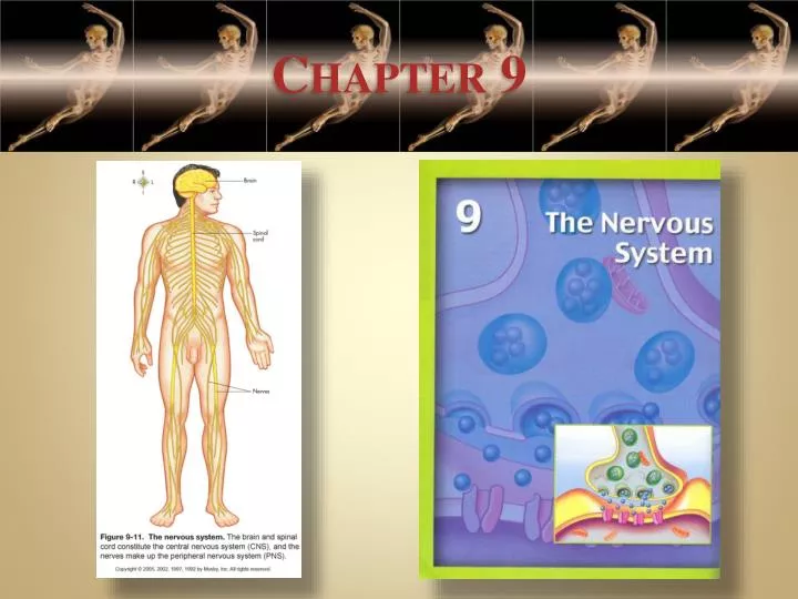
Central Nervous System(Figure 9-11) Page(s) 275
Central Nervous System • Divisions of the brain(Figure 9-12 and Table 9-1) • Brainstem • Consists of three parts of brain; named in ascending order, they are the medulla oblongata, pons, and midbrain • Structure—white matter with bits of gray matter scattered through it • Function—gray matter in the brainstem functions as reflex centers (for example, for heartbeat, respirations, and blood vessel diameter); sensory tracts in the brainstem conduct impulses to the higher parts of the brain; motor tracts conduct from the higher parts of the brain to the spinal cord Page(s) 274-275
Central Nervous System • Diencephalon • Structure and function of the hypothalamus • Consists mainly of the posterior pituitary gland, pituitary stalk, and gray matter • Acts as the major center for controlling the ANS; therefore, helps control the functioning of most internal organs • Controls hormone secretion by anterior and posterior pituitary glands; therefore it indirectly helps control hormone secretion by most other endocrine glands • Contains centers for controlling appetite, wakefulness, pleasure, etc. Page(s) 275-277
Central Nervous System • Structure and function of the thalamus • Dumbbell-shaped mass of gray matter in each cerebral hemisphere • Relays sensory impulses to cerebral cortex sensory areas • In some way produces the emotions of pleasantness or unpleasantness associated with sensations Page(s) 277
Central Nervous System • Cerebellum • Second largest part of the human brain • Helps control muscle contractions to produce coordinated movements so that we can maintain balance, move smoothly, and sustain normal postures • Recent evidence shows the cerebellum may also have wider coordinating effects, assisting the cerebrum and other regions of the brain Page(s) 277-278
Central Nervous System • Cerebrum • Largest part of the human brain • Outer layer of gray matter is the cerebral cortex; made up of lobes; composed mainly of dendrites and cell bodies of neurons • Interior of the cerebrum composed mainly of white matter (that is nerve fibers arranged in bundles called tracts) • Functions of the cerebrum—mental processes of all types, including sensations, consciousness, memory, and voluntary control of movements (Figure 9-13) Page(s) 278
Central Nervous System • Brain disorders • Destruction of brain tissue • Cerebrovascular accident (CVA)—hemorrhage from or cessation of blood flow through cerebral blood vessels; a “stroke” • Cerebral palsy—condition in which damage to motor control areas of the brain before, during, or shortly after birth causes paralysis (usually spastic) of one or more limbs Page(s) 278-280
Central Nervous System • Dementia—syndrome that includes progressive loss of memory, shortened attention span, personality changes, reduced intellectual capacity, and motor control deficit • Alzheimer disease (AD)—brain disorder of the middle and late adult years characterized by dementia • Huntington disease (HD)—inherited disorder characterized by chorea (purposeless movement) progressing to severe dementia • HIV (also causes AIDS) can infect neurons and thus cause dementia Page(s) 280-282
Central Nervous System • Seizure disorders • Seizure—sudden burst of abnormal neuron activity that results in temporary changes in brain function • Epilepsy—many forms, all characterized by recurring seizures • Electroencephalogram—graphic representation of voltage changes in the brain used to evaluate brain activity (Figure 9-14) Page(s) 282
Central Nervous System Spinal cord (Figure 9-15) Page(s) 284
Central Nervous System • Outer part is composed of white matter made up of many bundles of axons called tracts; interior composed of gray matter made up mainly of neuron dendrites and cell bodies (Figure 9-16) • Functions as the center for all spinal cord reflexes; sensory tracts conduct impulses to the brain, and motor tracts conduct impulses from the brain Page(s) 282-285 Spinal cord
Central Nervous System • Coverings and fluid spaces of the brain and spinal cord • Coverings • Cranial bones and vertebrae • Cerebral and spinal meninges—the dura mater, arachnoid mater, and the pia mater (Figure 9-17) Page(s) 285-286
Central Nervous System • Coverings and fluid spaces of the brain and spinal cord • Fluid spaces—subarachnoid spaces of meninges, central canal inside cord, and ventricles in brain (Figure 9-18 to 9-20) Page(s) 285-286
Peripheral Nervous System • Cranial nerves (Figure 9-21 and Table 9-2) • Twelve pairs—attached to undersurface of the brain Page(s) 286-289 • Connect brain with the neck and structures in the thorax and abdomen
Peripheral Nervous System • Spinal nerves • Structure—contain dendrites of sensory neurons and axons of motor neurons • Function—conduct impulses necessary for sensations and voluntary movements (Figure 9-22) Page(s) 290-292
Peripheral Nervous System • Peripheral nerve disorders • Neuritis—general term referring to nerve inflammation • Sciatica is inflammation of the sciatic nerve that innervates the legs • Neuralgia, or muscle pain, often accompanies neuritis Page(s) 292
Peripheral Nervous System • Trigeminal neuralgia—recurring episodes of stabbing pain along one or more branches of the trigeminal (fifth cranial) nerve in the head • Bell’s palsy—paralysis of facial features resulting from damage to the facial (seventh cranial) nerve Page(s) 292
Peripheral Nervous System • Herpes zoster or shingles (Figure 9-23) • Viral infection caused by chickenpox virus that has invaded the dorsal root ganglion and remained dormant until an episode of shingles • Usually affects a single dermatome, producing characteristic painful plaques or vesicles Page(s) 292-293