Download
1 / 53
530 likes | 550 Vues
Diabetic Foot Management DR.Ghazi Saeid. Introduction. Diabetes is one of the leading causes of chronic disease and limb loss worldwide, currently affecting 382 million people . It is predicted that by 2035, the number of reported diabetes cases will soar to 592 million .

E N D
Introduction • Diabetes is one of the leading causes of chronic disease and limb loss worldwide, currently affecting 382 million people. • It is predicted that by 2035, the number of reported diabetes cases will soar to 592 million. • Every year, >1 million people with diabetes suffer limb loss as a result of diabetes. • Every 20 seconds, an amputation occurs in the world.
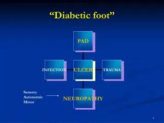
Risk factors for ulceration : • Neuropathy • PAD • Foot deformity • Limited ankle range of motion • High plantar foot pressures • Minor trauma • Previous ulceration or amputation • Visual impairment • At least one-quarter of these ulcers will not heal, and up to 28% may result in some form of amputation.
Prevention of diabetic foot ulceration • Off-loading DFUs • Diagnosis of diabetic foot osteomyelitis (DFO) • Wound care for DFUs • Peripheral arterial disease (PAD) and the DFU
Recommendation 1. We recommend that patients with diabetes undergo annual interval foot inspections by physicians (MD, DO, DPM) or advanced practice providers with training in foot care (Grade 1C). • Recommendation 2. We recommend that foot examination include testing for peripheral neuropathy using the Semmes-Weinstein test (Grade 1B).
The patient should be questioned about leg discomfort. • What is the sensation felt? – Burning, numbness, or tingling (2 points); fatigue, cramping, or aching (1 point). Maximum is 2 points. • What is the location of symptoms? – Feet (2 points); calves (1 point); elsewhere (no points). Maximum is 2 points. • Have the symptoms ever awoken you at night? – Yes (1 point). • What is the timing of symptoms? – Worse at night (2 points); present day and night (1 point); present only during the day (no points). Maximum is 2 points. • How are symptoms relieved? – Walking around (2 points); standing (1 point); sitting or lying or no relief (no points). Maximum is 2 points. • 0 to 2 – Normal • 3 to 4 – Mild • 5 to 6 – Moderate • 7 to 9 – Severe
Recommendation 3. We recommend education of the patients and their families about preventive foot care (Grade 1C).
Recommendation 4. • a. We suggest against the routine use of specialized therapeutic footwear in average-risk diabetic patients (Grade 2C). • b. We recommend using custom therapeutic footwear in high-risk diabetic patients, including those with significant neuropathy, foot deformities, or previous amputation (Grade 1B).
Recommendation 5. We suggest adequate glycemic control (hemoglobin A1c < 7% with strategies to minimize hypoglycemia) to reduce the incidence of DFUs and infections, with subsequent risk of amputation (Grade 2B). • hemoglobin A1c may be a useful marker for DFU healing; in a study of 183 patients with DFU, every increase of 1% in glycosylated hemoglobin decreases wound healing rate by 0.028 cm/d.
Recommendation 6. We recommend against prophylactic arterial revascularization to prevent DFU (Grade 1C). • For patients with diabetes and tissue loss in the setting of significant PAD, revascularization to prevent limb loss is well justified (Grade 1B).
Prevention of diabetic foot ulceration • Off-loading DFUs • Diagnosis of diabetic foot osteomyelitis (DFO) • Wound care for DFUs • Peripheral arterial disease (PAD) and the DFU
Recommendation 1. In patients with plantar DFU, we recommend off-loading with a total contact cast (TCC) or irremovable fixed ankle walking boot (Grade 1B). • Recommendation 2. In patients with DFU requiring frequent dressing changes, we suggest off-loading using a removable cast walker (RCW) as an alternative to TCC and irremovable fixed ankle walking boot (Grade2C).We suggest against using postoperative shoes or standard or customary footwear for off-loading plantar DFUs (Grade 2C).
Recommendation 3. In patients with non plantar wounds, we recommend using any modality that relieves pressure at the site of the ulcer, such as a surgical sandal or heel relief shoe (Grade 1C). • Recommendation 4. In high-risk patients with healed DFU (including those with a prior history of DFU, partial foot amputation, or Charcot foot), we recommend wearing specific therapeutic footwear with pressure-relieving insoles to aid in prevention of new or recurrent foot ulcers (Grade 1C).
Prevention of diabetic foot ulceration • Off-loading DFUs • Diagnosis of diabetic foot osteomyelitis (DFO) • Wound care for DFUs • Peripheral arterial disease (PAD) and the DFU
Risk factors for the development of Diabetic Foot Infections : • Neuropathy • Vasculopathy • Immunopathy • Diabetic Foot Osteomilitismay be present in up to 20% of mild to moderate infections and in 50% to 60% of severely infected wounds.
Wagner classification • Grade 0 – No ulcer in a high riskfoot • Grade 1 – Superficial ulcer involving the full skin thickness but not underlying tissues • Grade 2 – Deep ulcer, penetrating down to ligaments and muscle, but no bone involvement or abscess formation • Grade 3 – Deep ulcer with cellulitis or abscess formation, often with osteomyelitis • Grade 4 – Localized gangrene • Grade 5 – Extensive gangrene involving the whole foot
University of Texas system • Grade: • Grade 0: Pre orpost ulcerative (Stage A-D) • Grade 1: Full thicknessulcer not involving tendon, capsule, or bone (Stage A-D) • Grade 2: Tendon or capsular involvement without bone palpable (Stage A-D) • Grade 3: Probes to bone (Stage A-D) • Stage: • A: Non infected • B: Infected • C: Ischemic • D: Infected and ischemic
Recommendation 1. In patients with a DFI with an open wound, we suggest doing a probe to bone (PTB) test to aid in diagnosis (Grade 2C). (sensitivity 60%and specificity 91%, positive predictive value 89%.) • Recommendation 2. In all patients presenting with a new DFI, we suggest that serial plain radiographs of the affected foot be obtained to look for bone abnormalities (deformity, destruction) as well as soft tissue gas and radiopaque foreign bodies (Grade 2C). (sensitivity 54%and specificity 68%)
Recommendation 3. For those patients who require additional (ie, more sensitive or specific) imaging, particularly when soft tissue abscess is suspected or the diagnosis of osteomyelitis remains uncertain, we recommend using MRI as the study of choice. MRI is a valuable tool for diagnosis of osteomyelitis if the plain film is not useful (Grade 1B). (sensitivity 90%and specificity 79%)
Recommendation 4. In patients with suspected DFO for whom MRI is contraindicated or unavailable, we suggest a leukocyte or antigranulocyte scan, preferably combined with a bone scan as the best alternative (Grade 2B). (sensitivity 81%and specificity 28%) • Recommendation 5. In patients at high risk for DFO, we recommend that the diagnosis is most definitively established by the combined findings on bone culture and histology (Grade 1C). When bone is débrided to treat osteomyelitis, we recommend sending a sample for culture and histology (Grade 1C).
Recommendation 6. For patients not undergoing bone débridement, we suggest that clinicians consider obtaining a diagnostic bone biopsy when faced with diagnostic uncertainty, inadequate culture information, or failure of response to empirical treatment (Grade 2C).
Prevention of diabetic foot ulceration • Off-loading DFUs • Diagnosis of diabetic foot osteomyelitis (DFO) • Wound care for DFUs • Peripheral arterial disease (PAD) and the DFU
Recommendation 1. We recommend frequent evaluation at 1- to 4-week intervals with measurements of diabetic foot wounds to monitor reduction of wound size and healing progress (Grade 1C). • Wound area reduction of 10% to 15% per week or >= 50% area reduction in 4 weeks results in increased likelihood of healing with decreased complications of infection and amputation.
Recommendation 1.1 : We recommend evaluation for infection on initial presentation of all diabetic foot wounds, with initial sharp débridement of all infected diabetic ulcers, and urgent surgical intervention for foot infections involving abscess, gas, or necrotizing fasciitis (Grade 1B). • Recommendation 1.2 : We suggest that treatment of DFIs should follow the most current guidelines published by the IDSA (Ungraded).
Recommendation 2. We recommend use of dressing products that maintain a moist wound bed, control exudate, and avoid maceration of surrounding intact skin for diabetic foot wounds (Grade 1B). • Optimal wound care provides moist coverage, absorption of exudate, autolytic débridement, prevention of infection, and promotion of granulation. Non adherent dressings that protect the wound bed are standard treatment for most wounds.
Key Massage • Currently there is no research evidence to suggest that any type of hydrocolloid wound dressing is more effective in healing diabetic foot ulcers than other types of dressing or a topical cream containing plant extracts. Decision makers may wish to consider aspects such as dressing cost and the wound management properties offered by each dressing type e.g. exudate management.
Key Massage • Currently there is no research evidence to suggest that foam wound dressings are more effective in healing foot ulcers in people with diabetes than other types of dressing however all trials in this field are very small. Decision makers may wish to consider aspects such as dressing cost and the wound management properties offered by each dressing type e.g. exudate management.
Key Massage • Currently there is no research evidence to suggest that alginate wound dressings are more effective in healing foot ulcers in people with diabetes than other types of dressing however many trials in this field are very small. Decision makers may wish to consider aspects such as dressing cost and the wound management properties offered by each dressing type e.g. exudate management.
Key Massage • There is some evidence to suggest that hydrogel dressings are more effective in healing (lower grade) diabetic foot ulcers than basic wound contact dressings however this finding is uncertain due to risk of bias in the original studies. There is currently no research evidence to suggest that hydrogel is more effective than larval therapy or platelet-derived growth factors in healing diabetic foot ulcers, nor that one brand of hydrogel is more effective than another in ulcer healing. No RCTs comparing hydrogel dressings with other advanced dressing types were found.
Recommendation 3. We recommend sharp débridement of all devitalized tissue and surrounding callus material from diabetic foot ulcerations at 1- to 4-week intervals (Grade 1B). • Recommendation 4. Considering lack of evidence for superiority of any given débridement technique, we suggest initial sharp débridement with subsequent choice of débridementmethod based on clinical context, availability of expertise and supplies, patient tolerance and preference, and cost-effectiveness (Grade 2C).
Recommendation 5. For DFUs that fail to demonstrate improvement (>50% wound area reduction) after a minimum of 4 weeks of standard wound therapy, we recommend adjunctive wound therapy options. These include negative pressure therapy, biologics (PDGF, living cellular therapy, extracellular matrix products, amnionic membrane products), and hyperbaric oxygen therapy. Choice of adjuvant therapy is based on clinical findings, availability of therapy, and cost-effectiveness; there is no recommendation on ordering of therapy choice. Re-evaluation of vascular status, infection control, and off-loading is recommended to ensure optimization before initiation of adjunctive wound therapy (Grade 1B).
Recommendation 6. We suggest the use of negative pressure wound therapy (NPWT) for chronic diabetic foot wounds that do not demonstrate expected healing progression with standard or advanced wound dressings after 4 to 8 weeks of therapy (Grade 2B). • Recommendation 7. We suggest consideration of the use of PDGF (becaplermin) for the treatment of DFUs that are recalcitrant to standard therapy (Grade 2B).
Recommendation 8. We suggest consideration of living cellular therapy using a bilayeredkeratinocyte/fibroblast construct or a fibroblast-seeded matrix for treatment of DFUs when recalcitrant to standard therapy (Grade 2B). • Recommendation 9. We suggest consideration of the use of extracellular matrix products employing acellularhuman dermis or porcine small intestinal submucosal tissue as an adjunctive therapy for DFUs when recalcitrant to standard therapy (Grade 2C).
Recommendation 10. In patients with DFU that fails to respond to 4 to 6 weeks of conservative management, we suggest hyperbaric oxygen therapy (Grade 2B).
Prevention of diabetic foot ulceration • Off-loading DFUs • Diagnosis of diabetic foot osteomyelitis (DFO) • Wound care for DFUs • Peripheral arterial disease (PAD) and the DFU
Recommendation 1.1. We suggest that patients with diabetes have ABI measurements performed when they reach 50 years of age (Grade 2C). • Recommendation 1.2. We suggest that patients with diabetes who have a prior history of DFU, prior abnormal vascular examination, prior intervention for peripheral vascular disease, or known atherosclerotic cardiovascular disease (eg, coronary, cerebral, or renal) have an annual examination of the lower extremities and feet including ABI and toe pressures (Grade 2C).
Recommendation 2. We recommend that patients with DFU have pedal perfusion assessed by ABI, ankle and pedal Doppler arterial waveforms, and either toe systolic pressure or transcutaneous oxygen pressure (TcPO2) annually (Grade 1B).