Download
1 / 44
1.61k likes | 4.53k Vues
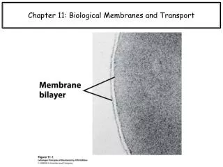
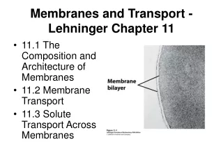
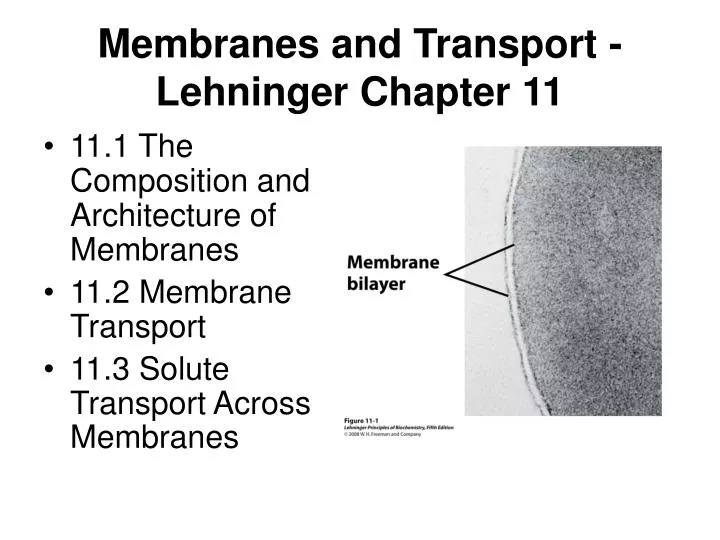
Membranes and Transport - Lehninger Chapter 11. 11.1 The Composition and Architecture of Membranes 11.2 Membrane Transport 11.3 Solute Transport Across Membranes. 11.1 The Composition and Architecture of Membranes. Membranes contain specialized lipids and proteins Membranes are fluid mosaics

E N D
Membranes and Transport - Lehninger Chapter 11 • 11.1 The Composition and Architecture of Membranes • 11.2 Membrane Transport • 11.3 Solute Transport Across Membranes
11.1 The Composition and Architecture of Membranes • Membranes contain specialized lipids and proteins • Membranes are fluid mosaics • Lipid Bilayer Structure • Peripheral Membrane Proteins • Integral membrane proteins • Sequence predictions of Membrane spanning domains • Lipid anchors
Membranes contain specialized lipids and proteins • Proteins 30-70% • Phospholipids 7-40% • Sterols 0-25% • Specialized membranes More than 90% Rhodopsin in photoreceptor disc membrane Protein rich mitochondrial membranes Transport optimized Red Blood Cell membrane
Membranes are fluid mosaics • Proteins/specialized structures as the tiles • Lipids as the mortar • Components are constrained to a plane but can diffuse laterally • Individual components diffuse, associate, dissociate in 2D
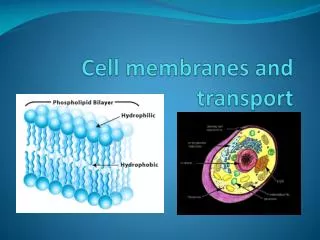
Lipid Bilayer Structure • Lipids assemble to segregate polar end nonpolar substituents • Micelles are globular • Exterior polar head group, Interior hydrophobic tail • Favored by single acyl chains • Vesicles or liposomes have an internal aqueous compartments • Inner and Outer leaflets • Bilayers are locally planar structures • Inner and Outer leaflets are asymmetrical • Bio membranes are ~3 nm (30 Angstroms) thick
Membrane Proteins • Peripheral membrane proteins • Associate with the membrane surface (lipids or protein) • Can be dissociated by changes in solution conditions (salt, pH) • Integral membrane proteins • Interact with hydrophobic bilayer core • Membrane Spanning • Lipid Anchors
Integral membrane proteins • Specific structure and orientation • Topology and orientation can be probed in intact membranes Protease digestion Chemical reactivity (lysine, cysteine modifications) • Membrane spanning segments expose nonpolar surfaces to bilayer interior • Structures of a few transmembrane proteins reveal common helical membrane spanning elements
Sequence predictions of Membrane spanning domains • Hydropathy plots average hydrophobicity over N successive residues (N=7-20) alpha helix dimensions 3.6 residues/turn over 5.4 Angstroms Bilayer dimensions ~30 Angstroms / 5.4 = ~6 turns of helix or 21 AAs • Hydrophobic stretches of ~20 residues are often membrane spanning helices • 7-9 residues of (more extended) beta strands can span the membrane
Lipid anchors • Post translational modification of Amino acids with Fatty Acids -- Palmitate, Myristate Isoprenoids -- Farnesyl, geranyl (Inner leaflet) Sterols Glycosyl Phosphatidyl Inositol (GPI) - (Outer leaflet)
11.1 Summary • Membrane compartments • The fluid mosaic model • Peripheral and integral membrane proteins • Membrane spanning proteins • Asymmetry
11.2 Membrane Transport • Acyl Chains - order-disorder transition • Transbilayer transport by flippases • Lateral diffusion • Lipid Rafts • Caveolae • Cell adhesion proteins • Membrane fusion
Acyl Chains - order-disorder transition • Sterols and straight chains favor order • Double bonds and short chains favor disorder • Lipid composition is adjusted to maintain constant fluidity High temperature - more saturated FAs, sterols Low temperature - more unsaturated FAs, shorter chains
Transbilayer transport by flippases • Barriers to transbilayer lipid movement are high • Lipid biosynthesis on one side of a membrane is coupled to catalyzed transport
Lateral diffusion • Lipids and proteins can diffuse in 2D • Measured by Fluorescence Recovery After Photo-bleaching FRAP requires fluorescent tag on lipid or protein Photobleaching of a small area by intense light pulse makes a dark spot Recovery depends on diffusion of undamaged fluorophores to the bleached spot
Lateral diffusion may be limited by protein networks Cytoskeletal connections or membrane patches
Lipid Rafts • Glycosphingolipids cluster in the outer membrane • Cholesterol also enriched in Lipid Rafts • GPI, palmitoyl and myristoyl anchors on signaling proteins enriched
Caveolae • Caveolin is an integral membrane protein Binds to the inner membrane Dimer, Palmitoyl anchors Induces curvature • caveolae (as in cave) on the extracellular side • bulges on thy cytoplasmic side Caveolae seem to be the locus for signalling
Cell adhesion proteins - extracellular domains • Integrins - attach to the extracellular matrix Bind collagen and fibronectin • Cadherins - Homotypic association Side by side dimers on the same cell Interactions between adjacent cells • Selectins - bind to oligosaccharides Cell surface lectins
Membrane fusion • Budding and fusion are two sides of the same coin • Fusion 1. Recognition 2. Apposition 3. Disruption 4. Bilayer fusion
Neurotransmitter release due to vesicle fusion at gap junctions SNAP - NSF Attachment Protein SNARE - Soluble NSF Attachment protein REceptor
11.2 Summary • Order and fluidity • Leaflets are isolated except for catalyzed exchange • Lateral diffusion allows for assembly of lipid rafts • Caveolae as signalling centers • Integrins, adhesins • Membrane fusion
11.3 Solute Transport Across Membranes • Passive transport • Transport super-families • Erythrocyte glucose transporter • Chloride-Bicarbonate Exchange • Active Transport • P-type ATPases • F-type ATPases • ABC transporter • Ion Gradients • Aquaporins • Ion selective channels • Sodium Channels and Nerve function • Acetycholine receptor - a ligand gated ion channel • Ion channel defects and inhibitors
11.3 Solute Transport Across Membranes Simple Diffusion Facilitated Diffusion Pores, Channels Ionophores Active Transport Direct Coupled
Energetics of Transport - Chemical Potential • Free energy defines the driving force • Transport of electro-neutral species is governed by chemical potential differences The chemical potential μ is made up of a standard state potential μ0 and a concentration dependent term μ = μ0 + RTlnC The free energy difference between two solutions that differ only in the concentration of one component is the difference between the chemical potentials ΔG = μ1 - μ2 = (μ0 + RTlnC1) - (μ0 + RTlnC2) = RTln(C1/C2)
For the free energy difference between the inside and outside • ΔG = RTln(Cin /Cout) • This is the driving force (or cost) of the transport reaction
Energetics of Transport - Electrical Potential • The energetic driving force (or cost) of transporting a charged object across an electric field ΔGel = Z F Δψ Δψ is the electrical potential in volts Z is the charge on the ion F is the Faraday constant 96,480 J/(V mole)
Energetics of Transport - Electro-chemical Potential • ΔG = RTln(Cinn/Cout) + ZF Δψ • If you forget the signs, remember: Free energy of a spontaneous reaction is negative Diffusion is spontaneous from high concentration to low Electrical transport is spontaneous for charges (Z) with signs opposite the potential difference (Δψ)
Passive transport - facilitated diffusion by proteins • Simple diffusion Rate determined by lipid/aqueous solubility Driving force is the sum of • Simple chemical potential and • electrochemical potential Not saturable (no Vmax) • Facilitated Diffusion - "passive transport” Transporters or Permeases are proteins Directionality determined by concentration and electrochemical gradients
Transport superfamilies • Carriers • Bind specific ligands • Catalyze transport across the membrane • In a sense they are enzymes • Transport is saturable • Channels • Less specific (often size specific) • Can be fluid filled • Transport may not be easily saturable
Erythrocyte glucose transporter - Uniporter • GlUT1 transports glucose into red blood cells • Specific for glucose, over other sugars
Michaelis Menten Kinetics • E + S <==> ES --> E + P • E + Sout <==> { ESo <==> ESi } --> E + Sin • Transition state is now conformational change of transporter • v0 = (Vmax [S]out) / (KM + [S]out) • Again v0 is the initial rate, - applies only when [S]out = constant, [S]in is negligible • If [S]in is not negligible, back reaction must be considered • E + Sout <==> { ESo <==> ESi } <==> E + Sin
Chloride-Bicarbonate Exchange - Antiporter • Chloride and Bicarbonate • carry the same charge • move in opposite directions - ping pong mechanism • maintain electro-neutrality • Cl-in + E <=> E + Cl-out • HCO3-out + E <=> E + HCO3-in
Active Transport • Energy coupling can transport against a concentration gradient • Primary • Transport is coupled to a chemical process (ATP hydrolysis) • Secondary • Transport is coupled to a favorable transport process
P-type ATPases - Active Transport • Transport phosphate coupled to ATP hydrolysis • Inhibited by vanadate • Na+K+ ATPase • 2K+out + 3Na+in + ATP --> 2K+in + 3Na+out + ADP + Pi • Net charge (+1) transfer out results in a -50-70 mV membrane potential • Energetically costly but membrane potential essential for action potential and other processes
F-type ATPases - Proton Gradients <==> ATP • Can either use ATP to pump protons or proton gradients to make ATP
ABC transporters - homologous family • classified by sequence and structure - not by function • ATP dependent transport • Multidrug resistance transporter pumps out foreign compounds • The chloride channel CFTR responsible for cystic fibrosis • Flippases for transbilayer lipid transport
Ion Gradients - Na+ or H+ can drive secondary transport • lac permease - bacterial lactose proton symport • Active transport of Lactose depends on maintenance of proton gradient
Na+- Glucose Symport in human intestine • 2 Na+out + Glucoseout --> 2 Na+in + Glucosein • Combination of sodium chemical potential and membrane potential • provide driving force for ~9000 fold concentration [Glucose]in/[Glucose]out
Aquaporins • Allow passive transport of water • Respond to changes in osmotic pressure
Ion selective channels • Ligand gated • Acetylcholine Receptor • Neuromuscular junction • Voltage Gated • K+ Channel
Ion channel measurements • Patch Clamping can measure the characteristics of a single channel • Glass micropipette can be used to capture a "patch" of membrane with one or more channels • Patch detached from the cell - seals the pipet opening
Solute Transport Summary • Passive and Active Transport • Carriers • Electrochemical Driving Forces • Coupled Transport • Ion Channels