Download
1 / 1
30 likes | 356 Vues
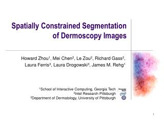
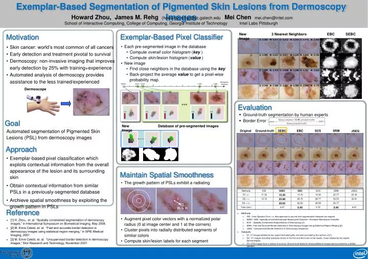
r. {R, G, B}. Exemplar-Based Segmentation of Pigmented Skin Lesions from Dermoscopy Images. Howard Zhou, James M. Rehg {howardz,rehg}@cc.gatech.edu School of Interactive Computing, College of Computing, Georgia Institute of Technology. Mei Chen mei.chen@intel.com Intel Labs Pittsburgh.

E N D
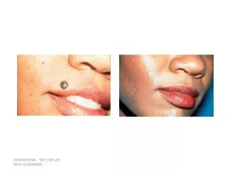
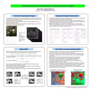
r {R, G, B} Exemplar-Based Segmentation of Pigmented Skin Lesions from Dermoscopy Images Howard Zhou, James M. Rehg {howardz,rehg}@cc.gatech.eduSchool of Interactive Computing, College of Computing, Georgia Institute of Technology Mei Chen mei.chen@intel.comIntel Labs Pittsburgh New Image 3 Nearest Neighbors EBC SEBC Exemplar-Based Pixel Classifier Motivation • Skin cancer: world’s most common of all cancers • Early detection and treatment pivotal to survival • Dermoscopy: non-invasive imaging that improves early detection by 25% with training+experience • Automated analysis of dermoscopy provides assistance to the less trained/experienced • Each pre-segmented image in the database • Compute overall color histogram (key ) • Compute skin/lesion histogram (value ) • New image • Find close neighbors in the database using the key • Back-project the average value to get a pixel-wise probability map. Original Ground-truth SEBC EBC SCS SRM JSEG Dermoscope Evaluation • Ground-truth segmentation by human experts • Border Error : Goal New Image Database of pre-segmented images Automated segmentation of Pigmented Skin Lesions (PSL) from dermoscopy images Approach • Exemplar-based pixel classification which exploits contextual information from the overall appearance of the lesion and its surrounding skin • Obtain contextual information from similar PSLs in a previously-segmented database • Archieve spatial smoothness by exploiting the growth pattern in PSLs Maintain Spatial Smoothness • The growth pattern of PSLs exhibit a radiating appearance Reference • Methods • IOE : Intra-Operator Error, i.e. discrepancies in ground-truth segmentation between two experts • SEBC / EBC: Spatially-smoothed Exemplar-Based pixel Classifier / Exemplar-Based pixel Classifier • SCS : Spatially Constrained Segmentation of Dermoscopy [1] • SRM : Fast and Accurate Border Detection in Dermoscopy Images Using Statistical Region Merging [2] • JSEG : Unsupervised Border Detection in Dermoscopy Images [3] • Datasets • D1: 67 images labeled by two expert dermatologists, and was provided by the authors of [1]. • D2: 111 images including examples shown in the first and third rows of the results. It was labeled by two expert dermatologists. • D3: 2159 images from a variety of sources. Ground truth labels for these additional images were provided by a skilled operator. • Augment pixel color vectors with a normalized polar radius (0 at image center and 1 at the corners) • Cluster pixels into radially distributed segments of similar colors • Compute skin/lesion labels for each segment • [1] H. Zhou, et. al. “Spatially constrained segmentation of dermoscopy images,” in International Symposium on Biomedical Imaging, May 2008. • [2] M. Emre Celebi, et. al. “Fast and accurate border detection in dermoscopy images using statistical region merging,” in SPIE Medical Imaging, 2007. • [3] M. Emre Celebi, et. al. “Unsupervised border detection in dermoscopy images,” Skin Research and Technology, November 2007.