Download
1 / 39
390 likes | 494 Vues
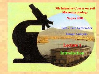
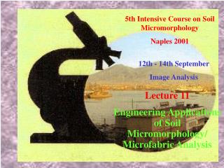
5th Intensive Course on Soil Micromorphology Naples 2001. 12th - 14th September Image Analysis. Lecture 9 Grey-Level Morphology and Multi-Spectral Methods. 5th Intensive Course on Soil Micromorphology - Naples 2001 Image Analysis - Lecture 9: Grey-Level Morphology. Part 1.

E N D
5th Intensive Course on Soil Micromorphology Naples 2001 12th - 14th September Image Analysis Lecture 9 Grey-Level Morphology and Multi-Spectral Methods
5th Intensive Course on Soil Micromorphology - Naples 2001 Image Analysis - Lecture 9: Grey-Level Morphology Part 1 Extension of Binary Morphology to Grey-Level Images avoids need to segment images Part 2 • Multi-Spectral Methods for Segmentation/Classification • useful where X-ray spectra of different colour information (e.g. RED/GREEN/BLUE/ U-V) information is available
5th Intensive Course on Soil Micromorphology - Naples 2001 Image Analysis - Lecture 9: Grey-Level Morphology Binary Morphology requires • Segmentation (usually by thresholding) and attendant problems • Erosion involves stripping pixels from edge of foreground areas according to selected criteria • Dilation involves adding pixels to foreground areas • Opening involves one cycle of erosion followed by one cycle or dilation • roughness aspects of feature are not recovered, no are particles smaller than 2 pixels • Closing is the reverse of Opening.
5th Intensive Course on Soil Micromorphology - Naples 2001 Image Analysis - Lecture 9: Grey-Level Morphology • Grey Level Morphology • attempts to solve problems of BINARY MORPHOLOGY • by removing need for thresholding • Grey Level Erosion replaces all intensities within a given • mask area by the minimum value in that area • Grey Level Dilation replaces all intensities within a given • mask area by the maximum value in that area • A grey level opening involves an erosion and a dilation phase • As with binary morphology, roughness is lost and features tend to become rounded until they finally disappear
5th Intensive Course on Soil Micromorphology - Naples 2001 Image Analysis - Lecture 9: Grey-Level Morphology Representation of binary morphology for feature sizing
5th Intensive Course on Soil Micromorphology - Naples 2001 Image Analysis - Lecture 9: Grey-Level Morphology Schematic of Intensity Profile along a line
5th Intensive Course on Soil Micromorphology - Naples 2001 Image Analysis - Lecture 9: Grey-Level Morphology Start of Erosion along line
5th Intensive Course on Soil Micromorphology - Naples 2001 Image Analysis - Lecture 9: Grey-Level Morphology Intensity lost after grey-level erosion (blue)
5th Intensive Course on Soil Micromorphology - Naples 2001 Image Analysis - Lecture 9: Grey-Level Morphology Intensity lost after grey-level erosion followed by dilation Blue: Intensity lost: Green: Intensity recovered in dilation
5th Intensive Course on Soil Micromorphology - Naples 2001 Image Analysis - Lecture 9: Grey-Level Morphology Intensity lost after grey-level erosion of diameter 5 Cyan: New Intensity lost
5th Intensive Course on Soil Micromorphology - Naples 2001 Image Analysis - Lecture 9: Grey-Level Morphology Intensity lost after grey-level erosion followed by dilation (diameter 5) Blue/ Cyan: Intensity lost: Green: Intensity recovered in dilation
5th Intensive Course on Soil Micromorphology - Naples 2001 Image Analysis - Lecture 9: Grey-Level Morphology Intensity lost after grey-level erosion of diameter 7 Purple: New Intensity lost
5th Intensive Course on Soil Micromorphology - Naples 2001 Image Analysis - Lecture 9: Grey-Level Morphology Intensity lost after grey-level erosion followed by dilation (diameter 7) Blue/ Cyan/Purple: Intensity lost: Green: Intensity recovered in dilation
5th Intensive Course on Soil Micromorphology - Naples 2001 Image Analysis - Lecture 9: Grey-Level Morphology Effect of grey-level opening at different radii
5th Intensive Course on Soil Micromorphology - Naples 2001 Image Analysis - Lecture 9: Grey-Level Morphology a) Radius 9 pixels b) Radius 10 pixels c) Difference Image d) Complete particle loss
5th Intensive Course on Soil Micromorphology - Naples 2001 Image Analysis - Lecture 9: Grey-Level Morphology 1 2 3 4 Particle size analysis using grey-level morphology Core sample taken from estuary model. [photograph courtesy of J.Alexander]
5th Intensive Course on Soil Micromorphology - Naples 2001 Image Analysis - Lecture 9: Grey-Level Morphology Halimeda needles from Great Barrier Reef
5th Intensive Course on Soil Micromorphology - Naples 2001 Image Analysis - Lecture 9: Grey-Level Morphology Halimeda needles from Great Barrier Reef
5th Intensive Course on Soil Micromorphology - Naples 2001 Image Analysis - Lecture 9: Grey-Level Morphology Halimeda needles from Great Barrier Reef - partly covered by nanograins
5th Intensive Course on Soil Micromorphology - Naples 2001 Image Analysis - Lecture 9: Grey-Level Morphology Halimeda needles from Great Barrier Reef - partly covered by nanograins
5th Intensive Course on Soil Micromorphology - Naples 2001 Image Analysis - Lecture 9: Grey-Level Morphology Halimeda needles from Great Barrier Reef - fully covered by nanograins
5th Intensive Course on Soil Micromorphology - Naples 2001 Image Analysis - Lecture 9: Grey-Level Morphology Question: Are nanograins biological or chemical in origin? Evidence suggests nanograins increase in size with coverage - hence favouring chemical argument.
5th Intensive Course on Soil Micromorphology - Naples 2001 Image Analysis - Lecture 9: Grey-Level Morphology If particles lost at each radius are stored and finally added (B) - the resulting image should be comparable to original (A). Except:All particles are reduced to their equivalent circular diameter.
5th Intensive Course on Soil Micromorphology - Naples 2001 Image Analysis - Lecture 9: Multi-Spectral Analysis • Allows alternative methods for segmentation • Enables separation of different mineral classes. • Can be used in combination with Orientation Analysis as a combination method to overcome problem of large particles Multi-Spectral Analysis • Requirements: • Two or more images of same area at same magnification and pixel resolution and in exact registry. • Must be collected with different physical parameters - e.g. wavelength
5th Intensive Course on Soil Micromorphology - Naples 2001 Image Analysis - Lecture 9: Multi-Spectral Analysis Requirements continued: • Examples: • Optical Microscopy: • Red / Green / Blue images • UV. • Electron Microscopy: • Secondary Electron • Back Scattered Electron • Cathodoluminescence • X-Ray Maps.
5th Intensive Course on Soil Micromorphology - Naples 2001 Image Analysis - Lecture 9: Multi-Spectral Methods Multi-Spectral Methods • Require 2 or more different images of same area • must be in exact registry • e.g. Optical Microscope • RED/GREEN/BLUE/UV Or SE / BSE Image and CL or various X - Ray Maps in SEM
5th Intensive Course on Soil Micromorphology - Naples 2001 Image Analysis - Lecture 9: Multi-Spectral Methods Hong Kong Marine Clay from M1 unit approximately 1m above upper most palaeo-desiccated layer. BSE Image
5th Intensive Course on Soil Micromorphology - Naples 2001 Image Analysis - Lecture 9: Multi-Spectral Methods Hong Kong Marine Clay
5th Intensive Course on Soil Micromorphology - Naples 2001 Image Analysis - Lecture 9: Multi-Spectral Methods BSE Image X-Ray Maps
5th Intensive Course on Soil Micromorphology - Naples 2001 Image Analysis - Lecture 9: Multi-Spectral Methods From N images and Statistics from M classes Output segmented image may be obtained. Accuracy in segmentation relies on identification of suitable classes, and also sufficient classes
5th Intensive Course on Soil Micromorphology - Naples 2001 Image Analysis - Lecture 9: Multi-Spectral Methods Are these two particles the same material? Classification was set at 98% confidence and some post-processing was done to produce classified image.
5th Intensive Course on Soil Micromorphology - Naples 2001 Image Analysis - Lecture 9: Multi-Spectral Methods Procedure of segmentation is know as Mineral-Segmentation
5th Intensive Course on Soil Micromorphology - Naples 2001 Image Analysis - Lecture 9: Multi-Spectral Methods
5th Intensive Course on Soil Micromorphology - Naples 2001 Image Analysis - Lecture 9: Multi-Spectral Methods Particle Size Distribution for different mineral species
5th Intensive Course on Soil Micromorphology - Naples 2001 Image Analysis - Lecture 9: Multi-Spectral Methods Use Mineral Segmented image to generate binary masks. Binary Mask to assess orientation in matrix outside aggregate. Large mineral grains and voids are black as is aggregate. Binary Mask to assess orientation in matrix inside aggregate.
5th Intensive Course on Soil Micromorphology - Naples 2001 Image Analysis - Lecture 9: Multi-Spectral Methods Domain Segmentation of Matrix
5th Intensive Course on Soil Micromorphology - Naples 2001 Image Analysis - Lecture 9: Multi-Spectral Methods a) Matrix orientation c) Quartz grain orientation e) Weighted Quartz grain orientation b) Aggregate orientation d) Feldspar orientation f) Weighted Feldspar orientation Hong Kong Marine Clay
5th Intensive Course on Soil Micromorphology - Naples 2001 Image Analysis - Lecture 9: Multi-Spectral Methods Index of Anisotropy outside aggregate: 0.229 inside aggregate: 0.374 In both cases the predominant orientation is nearly vertical. Vertical direction in field.
5th Intensive Course on Soil Micromorphology - Naples 2001 Image Analysis - Lecture 9: Multi-Spectral Methods Concluding Remark on Multi-spectral Analysis. When does a particle warrant separate identification from matrix? - depends on pixel resolution/magnification. In supervised classification it is helpful to avoid “forced” classification as this will identify features / minerals which may have been missed. Some post-processing of image in needed following Mineral-Segmentation to remove noise etc.