Download
1 / 41
480 likes | 1.03k Vues
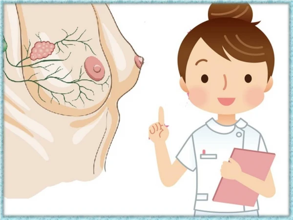
BREAST, CLINICAL. BY PROF. MOHAMED A. EL GHARBAWI Web Site: www.dr-elgharbawi.com E-mail: elgharma2@yahoo.com. EMBRYOLOGY, MILK LINE. CROSS SECTION OF THE BREAST. 1. CHEST WALL 2. PECTORALIS MUSCLE 3. LOBULES 4. NIPPLE 5. AREOLA 6. MILK DUCT 7. BREAST FAT 8. SKIN.

E N D
BREAST, CLINICAL BY PROF. MOHAMED A. EL GHARBAWI Web Site:www.dr-elgharbawi.com E-mail:elgharma2@yahoo.com
CROSS SECTION OF THE BREAST 1. CHEST WALL 2. PECTORALIS MUSCLE 3. LOBULES 4. NIPPLE 5. AREOLA 6. MILK DUCT 7. BREAST FAT 8. SKIN
CASE SENARIOHISTORY • Personal history :حميده محمد السيد40 years old femaleManual worker at Alnassagoon Al sharkeoon in Al AASHERLiving in BelbeesNo special habitsMarried with 4 living children • Main complaint:Lemon sized lump in left breast accidentally discovered before8 months • History of present illnessOnset: the lump was accidentally discovered during bathDuration: about 8 months agoCourse: lump gradually increasing in sizeThe lump became painful since 2 months
CASE SENARIOHISTORY • History of present illness (Cont.):No history of nipple dischargeNo change in the nipple appearanceNo sense of tension or pain during mensesNo weight lossNo history of bone pains, cough or expectorationNot on treatment for hypertension or diabetes
CASE SNARIOHISTORY • Past history:No history of taking oral contraceptive pills orhormonal therapyNo significant past medical historyAppendectomy before 20 years • Menstrual history:Menarche at age of 14 yearsRegular menstruationLast menstrual period before 15 days
CASE SNARIOHISTORY • Marital history:Married at age of 17 years5 pregnancies (4 vaginal deliveries + 1 abortion)First delivery at age of 18 years4 children2 girls (22 & 16 years)2 boys 20 & 8All are breast fed • Family history:2 brothers & 4 sistersNo history of breast cancer or other malignancies in the mother, grandma or sisters
CASE SNARIOGENERAL EXAMINATION • General look & Vital signs:162 cm long & 90 Kg (obese)ladyNo pallor No yellowish coloration of scleraNo cyanosisNo Clubbing of fingersNo generalized lymphadenopathyPulse: 88 per minuteBlood pressure: 145/ 90Temp. (oral): 37.3 C
CASE SNARIOGENERAL EXAMINATION • Other systems:Respiratory system: No signs of lung consolidation orpleural effusionAbdomen: No palpable liver, no other palpable abdominalmasses, no evidence of ascites.Skeletal system: No bone tenderness especially over the skull orspine Cardiovascular system: NAD • P/V & P/R: NAD
CASE SNARIOLOCAL EXAMINATION • INSPECTION:A. Sitting position & arms resting at sidesNormal similar contour of both breasts Both breasts are equal in size Both breasts are small and pendulous Both nipples are at the same levels No nipple retraction No apparent nipple discharge Slight elevation looking like a swelling in the upper outer quadrant of left breast Skin of breasts is looking normal with no signs of inflammation or ulceration No peau d’orange , no tethering or bickering No scars , no dilated veins
CASE SNARIOLOCAL EXAMINATION • INSPECTION:B. Sitting position & arms are raised above the head: No tethering or bickering of skin in both breasts
CASE SNARIOLOCAL EXAMINATION • PALPATION:A. Left breast (Site of main complaint):Superficial palpation:(To gain the confidence of the patient ) To check the skin temperature To test for tenderness Not warm Not tenderDeep palpation: (Verify what has been found by inspection)A single (Number) lemon (Shape)sized mass 2 by 2 cm (Size) is felt by bothtips of fingers & flat of hand at the upper outer quadrant (Site) ofleft breast
PALPATION SITTING& LYING Hands on
CASE SNARIOLOCAL EXAMINATION • PALPATION:A. Left breast (Site of main complaint):Edge: Ill defined (merge into the tissues)Consistency: Firm to hardMobility: Limited mobility, attached to surrounding breast tissue Attachments: Not attached to the skin over (You can pinch the skin over the mass) Not attached to the underlying pectoralis muscles or chest wall (when patient forward the mass moves forward + When the patient pushes her hands against her waist to contract the pectoralis muscle, the mass is not fixed)Pressure on the areola & nipple: No dischargePalpation while the patient is lying down: No nodularity
CASE SNARIOLOCAL EXAMINATION • PALPATION:B. Left Axilla: Two mobile, firm and non tender lymph nodes are palpable on the medial wall of the axilla. One cm each in sizeC. No palpable supraclavicular lymph nodesD. Right Breast & Axilla: NAD
CASE SENARIOPROVISIONAL DIAGNOSIS CARCINOMA OF LEFT BREAST, STAGE II T1 (Tumor size = 2 cm ) N1 (Ipsilateral mobile axillary lymph nodes) M0 ( No distant metastases)
CASE SENARIOINVESTIGATIONS • Confirmation of diagnosis: 1. Mammography 2. FNAC 3. May be True cut or excisional biopsy • For staging: 1. Mammography of other breast2. Abdominal US3. CXR4. X-ray to the bones where there is pain5. Bone Scan • Pre operative General evaluation: 1. CBC 2. Blood urea, creatinine & electrolytes 3. Liver function tests 4. ECG
CASE SENARIOTREATMENT • Surgery:Modified Radical Mastectomy • Histopathology & Test for Hormone receptors • Hormonal therapyRadiotherapyChemotherapyAccording to the results of pathology • Follow-up