Download
1 / 96
1.03k likes | 1.65k Vues
Chromosome Packaging, The Cell Cycle, Mitosis, & Cancer. Biology 11 Chapter 11. Part I. The discovery of Chromosomes & Understanding Chromosomal Organization in the Cell. Early Studies.

E N D
Chromosome Packaging, The Cell Cycle, Mitosis, & Cancer Biology 11 Chapter 11
Part I The discovery of Chromosomes & Understanding Chromosomal Organization in the Cell
Early Studies • As early as 1879 Walther Flemming was studying how “threadlike” structures change as salamander embryo divides • Greek mitos - thread • In 1888 Wilhelm Waldeyer coined the term chromosome “colored-body” to reference the thread like structure
Early Studies • As researchers watched rapidly growing plant cells at a root tip they realized that cell division is not continuous
Early Studies • There seemed to be two phases • Mitotic or M phase • Chromosomes can only be seen in this phase when they are highly condensed • Interphase or nondividing phase
Chromosome Organization During Interphase • Interphase is a time when cells are busy carrying out cellular business • Liver cell • Pancreatic cells • GI cell • In order to do their particular cellular work or cell “specialization” the certain chromosomal regions need to be decondensed
Chromosome Organization During Interphase • Decondensed chromosomes still must be bound to packaging proteins or the DNA would not “fit” into the nucleus • How is DNA is a cell packaged
Packaging DNA • DNA wraps around a conserved group of proteins known as histones • The 300 nm fiber attaches to scaffolding proteins • Actively transcribed regions are located on loops of the 300 nm fiber
Packaging DNA • The 300nm fiber coils into a increasing levels of compaction via mechanisms not fully understood • Particular genes are always located at the same position in a metaphase chromosome indicating that the packing steps are highly specific and precise
Histones • As a group these proteins are highly conserved between distantly related species • Histone H3 • Between a sea urchin and calf thymus it differs by only a single amino acid • Between a garden pea and calf thymus differ in only 4 amino acids • Significant deviations in amino acid sequence were selected against during evolution
Genes Chromosome Organization Looped Regions of 30nm fiber Scafold Proteins –MARs/SARs Looped regions can oscillate 10nm and 700nm fibers levels of compaction can change in a process known as chromatin remodeling Less compaction = transcription More compaction = no transcription
Euchromatin • Euchromatin – 10 nm to 300nm fiber • DNA which is not tightly packed • DNA bound to histones • Transcription occurs in euchromatin regions • Gene are “ON” • DNA is exposed to proteins and enzymes that facilitate gene expression
Heterochromatin • Heterochromatin • DNA which is tightly packaged • DNA bound to histones, but DNA histone complexes have increased folding • Varying levels of packing – repetitive sequences • Certain chromosomal regions are tightly packed even during interphase • During mitosis all chromosomes must maximally condense to facilitate organized chromosomal division
Accessing Genes in Euchromatin • Nontranscribed genes are more “exposed” to DNA enzymes Digests DNA
Accessing Genes in Euchromatin Question: Why is the 4.6kb band of DNA “gone” In the cells which express the globin gene? Answer: The DNA in the erythroblasts was Transcribed DNA and therefore not highly compacted and protected from digestion with DNAase.
Chromosome Organization During Interphase • Not all chromosomal areas are euchromatin even when the cell is in interphase • A liver cell will need to express different genes than a brain cell • Each cell will have different euchromatic regions • Each cell may have the same or different heterochromatic regions
Chromosome Organization During Interphase • Remember that the nucleus is a highly organized structure • Chromosome territories • Scaffold associated regions SARs • Matrix attachment regions MARs • Heterochromatin/Euchromatin boundaries • The currant model for chromosome organization in an interphase nucleus must take into account these and other factors
Chromosome Organization • Bottom line • Chromatin organization is complex and dynamic • However, during mitosis all chromosomes must become maximally condensed
Part II Mitosis & The Cell Cycle
Basic Concepts of Mitosis • Mitosis is a process of cell division which results in the production of two genetically identical daughter cells • Mitosis vs. cytokinesis • Mitosis is essentially a nuclear division • Cytokinesis is a division of the cytoplasm
Basic Concepts of Mitosis • Mitosis is responsible for three key events in cellular life • Growth • Wound repair • Reproduction • Asexual reproduction of single celled organisms • Asexual reproduction of cells in a multicellular organism • Somatic cells
Basic Concepts of Mitosis Haploid n 2n Diploid
The Discovery of the Cell Cycle • Chromosome moved to daughter cells • Each daughter cells ends up with the same number of chromosomes as the parent cells • Chromosome replication had to occur • When?? DNA Replication
The Discovery of the Cell Cycle This is an asynchronous culture Only cells which have just finished replicating DNA should be labeled
The Discovery of the Cell Cycle Labeled Cell Unlabeled Cell • The labeled cells should enter mitosis immediately if there is no delay between DNA replication and mitosis Condensed “mitotic” chromosomes seen Labeled cells in “interphase” no distinguishing chromosome
The Discovery of the Cell Cycle • Conclusion • There is a gap between replication and mitosis • Gap functions • Complete other requirements for cell division besides chromosome replication • Replication of organelles • Manufacture additional cytoplasm
The Discovery of the Cell Cycle • Subsequent research also showed that there was a gap between the exit from mitosis and DNA replication • All of this information led to the phases of a cell cycle
The Cell Cycle G0 G 1 S G 2
Basic Concepts of Cell Division • Most of your adult cells are not replicating their DNA and dividing • When you were just a single fertilized egg your cells were constantly dividing • Your cells had to specialize and grow • Bone • Liver cells • Skin cell
Basic Concepts of Cell Division • As a grown adult organism some cells are actively dividing • Skin • Wound healing and renewal • GI tract • Divide 2X per day to renew tissue lost during digestion • Liver • Divide 1X per year unless damaged • Unicellular Organism • Division is nutrient dependent
Basic Concepts of Cell Division • Even cells that are capable of replicating are not regularly going through cell division • Your cells are busy carrying out their specified functions and transcribing the necessary genes • Some cells rarely enter mitosis once they have matured • Post mitotic cells • Nerve cells • Muscle cells
Basic Concepts of Cell Division • Therefore, most of the time your cells are in what is known as a quiescent phase or “quite” phase • This phase is known as G0
Basic Concepts of Cell Division • Even when your cells divide they have to be “instructed to do so” • These instruction can be provided by • Hormones • Wounding
The Cell Cycle G0 G 1 S Concept Questions: What is the rate of this cycle in a Cancer Cell Embryonic Cell Liver Cell Liver tissue exposed to HepA Muscle Cell Human GI Cell G 2
G0 Phase where cells are performing their specialized tasks G1 Phase where cells have received the signal to grow and divide Proteins, enzymes and nucleotides are made which are needed to enter S phase S DNA is replicated G2 Proteins and enzymes are made which are necessary to complete M phase The Cell Cycle
M Phase • M Phase – Mitosis • This is the process by which the chromosomes actually separate • Usually a fast step • Typical human cells needs ~ 24 hours to divide • ~8 to 12 hours for DNA replication • ~11 hours for G phases • ~1 hour for Mitosis phase • These times are just examples; times can vary greatly from cell type to cell type
Part III Mechanisms of Mitosis
Mitosis • Mitosis is a nuclear division • One diploid cells becomes two diploid cells • Chromosomes duplicated exactly and separated Sister Chromatids One Duplicated Chromosome - exact genetic copy of each other
Basic Terms • Chromsome • Unduplicated chromosome • Sister chromatids • One duplicated chromosme • These are exact copies of each other One chromosome (unduplicated) Centromere one chromatid one chromatid One chromosome (duplicated) Two Sister Chromatids
Basic Terms - Centromere • Centromere is a DNA sequence found on each chromosome • Special centromere binding proteins associate with these sequences of DNA • Centromere binding proteins link together two sister chromosomes • When certain centromere binding proteins are cut apart the chromosomes can separate (division)
Basic Terms pole Centrioles Microtubule organizing center Not in plants Microtubules Dynamic motor (kinesin) proteins will associate with microtubules and pull the chromosomes apart Microtubules attaching to kinetochore Mitotic Spindle Spindle is only in animal cells Plants have microtubules but not spindle apparatus pole
Basic Terms - Kinetochore • Kinetochore • A protein structure at the centromere where spindle fibers (microtubules) attach to the sister chromatids of a replicated chromosome • Contains motor proteins
Two Sister Chromatids Kinetochore Protein Bridge Motor Proteins Centromere Binding Protein
Phases of Mitosis - Prophase • Prophase • Chromosomes condense • Centrioles have replicated • Mitotic spindle forms
Interphase in Animals This is a cell in interphase; a cell which is either is G1, S or G2. Since this is an embryo this cell is likely not in G0. The other circled cell is in _____________
Prophase in Animal Cells Chromosome Condense into threadlike structures
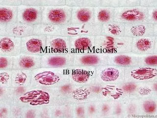
Prophase in Plant Cells Interphase Notice the nucleolus Three examples of Prophase
Phases of Mitosis – Prometaphase • Prometaphase • Nuclear envelope breaks down • Microtubules attach to kinetochore • One microtubule from each pole will attach to one duplicated chromosomes