Download
1 / 35
390 likes | 749 Vues
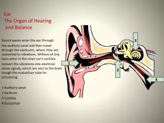
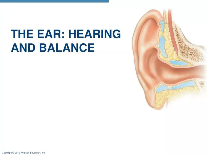
THE EAR: HEARING AND BALANCE. The Ear: Hearing and Balance. Three parts of the ear The External (outer) Ear Pinna- Composed of the Helix (rim) and lobule (earlobe) External acoustic canal (meatus)-short, curved tube leading to eardrum Lined with ceruminous glands

E N D
The Ear: Hearing and Balance • Three parts of the ear • The External (outer) Ear • Pinna- Composed of the Helix (rim) and lobule (earlobe) • External acoustic canal (meatus)-short, curved tube leading to eardrum • Lined with ceruminous glands • Tympanic membrane- eardrum; CT boundary btwn. external and middle ear; vibrates in response to sound
Middle ear Internal ear (labyrinth) External ear Auricle (pinna) Helix Lobule External acoustic meatus Tympanic membrane Pharyngotympanic (auditory) tube (a) The three regions of the ear Figure 15.25a
Middle Ear • The Middle Ear (tympanic cavity) • A small, air-filled, mucosa-lined cavity in the temporal bone; flanked laterally by the eardrum and medially by the oval and round windows • Three bones (ossicles)- Incus, Malleus,Stapes
Middle Ear • Two tiny skeletal muscles prevent damage due to large vibrations • Tensor tympani-arises from wall of pharyngotympanic tube and inserts on the malleus • Stapedius muscle-runs from posterior wall of middle ear to the stapes
Middle Ear • Pharyngotympanic (auditory) tubes • Connects middle ear to nasopharynx • Normally flattened closed; opens when yawning or swallowing • Equalizes pressure in the middle ear cavity with the external air pressure
Epitympanic recess Malleus Incus Superior Lateral Anterior View Pharyngotym- panic tube Tensor tympani muscle Tympanic membrane (medial view) Stapes Stapedius muscle Figure 15.26
Inner Ear • Consists of the bony (osseous) and membranous labyrinth • Bony labyrinth • Tortuous channels in the temporal bone • Filled with perilymph • Three regions: • Vestibule- • Contains two sacs • Saccule-continuous w/ the cochlear duct • Utricle-continuous w/ the semicircular ducts • Cochlea • Semicircular canals
The Inner Ear • Membranous Labyrinth • Series of membranous sacs and ducts contained w/in the bony labyrinth • Follows contours of bony labyrinth • Filled with endolymph
Superior vestibular ganglion Inferior vestibular ganglion Temporal bone Semicircular ducts in semicircular canals Facial nerve Vestibular nerve Anterior Posterior Lateral Cochlear nerve Cristae ampullares in the membranous ampullae Maculae Spiral organ (of Corti) Utricle in vestibule Cochlear duct in cochlea Saccule in vestibule Stapes in oval window Round window Figure 15.27
The Maculae and Static Equilibrium • Maculae- • Sensory receptors for static equilibrium (monitor the position of head in space, respond to linear acceleration) • One in each saccule wall and one in each utricle wall • Each maculae contains: • ET hair cells and supporting cells • Otolithic membrane- gel like membrane that overlies hair cells • Otoliths- calcium carbonate crystals; increase weight and its inertia
Otoliths Kinocilium Otolithic membrane Stereocilia Hair bundle Macula of utricle Macula of saccule Hair cells Supporting cells Vestibular nerve fibers Figure 15.34
Steps of linear movement • Hair cells are always releasing neurotransmitter • When hair cells bend towards kinocilium they depolarize, neurotransmitter release is increased • When hair cells bend away from kinocilium they hyperpolarize, slowing release of neurotransmitter • This change in neurotransmitter release informs the brain of the changing of the position of the head in space
Otolithic membrane Kinocilium Stereocilia Hyperpolarization Depolarization Receptor potential Nerve impulses generated in vestibular fiber When hairs bend toward the kinocilium, the hair cell depolarizes, exciting the nerve fiber, which generates more frequent action potentials. When hairs bend away from the kinocilium, the hair cell hyperpolarizes, inhibiting the nerve fiber, and decreasing the action potential frequency. Figure 15.35
The Crista Ampullaris and Dynamic Equilibrium • Dynamic Equilibrium • Detected by: • Crista Ampullaris – • One in the ampulla of each semicircular canal • Major stimuli are rotatory movements
The Crista Ampullaris and Dynamic Equilibrium • Semicircular Canals • Three canals are located in each ear: • Located in all three planes of space • Anterior, posterior and lateral • Endolymph-fills the semicircular ducts • Ampulla- swellling at end of semicircular duct • CristaAmpullaris • Composed of hair cells and supporting cells • Structure and function of the cristaampullaris is basically the same as the hair cells of cochlea and maculae • Cupula – gelled mass that cilia of hair cells are embedded in
Cupula Crista ampullaris Endolymph Hair bundle (kinocilium plus stereocilia) Hair cell Crista ampullaris Membranous labyrinth Supporting cell Fibers of vestibular nerve (a) Anatomy of a crista ampullaris in a semicircular canal Cupula (b) Scanning electron micrograph of a crista ampullaris (200x) Figure 15.36a–b
The Crista Ampullaris and Dynamic Equilibrium • Steps of Rotational Movement • At rest the cupula stands upright • During rotational acceleration, hair cells are bent, they depolarize and impulses reach the brain faster • As movement slows, endolymph keeps moving, cilia are bent in opposite direction causing hyperpolarization and reduction of impulses to brain
Section of ampulla, filled with endolymph Fibers of vestibular nerve Cupula Flow of endolymph At rest, the cupula stands upright. During rotational acceleration, endolymph moves inside the semicircular canals in the direction opposite the rotation (it lags behind due to inertia). Endolymph flow bends the cupula and excites the hair cells. As rotational movement slows, endolymph keeps moving in the direction of the rotation, bending the cupula in the opposite direction from acceleration and inhibiting the hair cells. (c) Movement of the cupula during rotational acceleration and deceleration Figure 15.36c
Equilibrium Pathway to the Brain • Vestibular nerve-Impulses travel to the vestibular nuclei in the brain stem or the cerebellum • Pathways are complex and poorly traced
Sound and the Cochlea • Sound is detected by: the cochlea • Cochlea • The cochlea is A spiral, conical, bony chamber and contains the cochlear duct • Cochlear duct- houses the spiral organ (of Corti) • Divides cochlea into three chambers: • Scala vestibuli-superior to cochlear duct (contains perilymph) • Scala tympani-inferior to cochlear duct; terminates at round window (contains perilymph) • Scala media (cochlear duct) -middle cavity; (contains endolymph)
Modiolus Cochlear nerve, division of the vestibulocochlear nerve (VIII) Spiral ganglion Osseous spiral lamina Vestibular membrane Cochlear duct (scala media) Helicotrema (a) Figure 15.28a
Vestibular membrane Osseous spiral lamina Tectorial membrane Spiral ganglion Scala vestibuli (contains perilymph) Cochlear duct (scala media; contains endolymph) Spiral organ (of Corti) Scala tympani (contains perilymph) Basilar membrane (b) Figure 15.28b
Sound and the Cochlea • Oval window-an opening on the medial wall of the middle ear (foot of stapes rests at oval window) • Round window-an opening on the medial wall of the middle ear (scala tympani terminates at round window) • Vestibular membrane-roof of cochlear duct that separates the scala media from scala vestibuli • Basilar membrane- fibrous floor of cochlear duct
Sound and the Cochlea • Organ of Corti • Runs through center of cochlea • Has hair cells and supporting cells • Tectorial membrane- gel-like mass that cilia of hair cells are embedded in • Bending of the cilia: excites hair cells
Tectorial membrane Inner hair cell Hairs (stereocilia) Afferent nerve fibers Outer hair cells Supporting cells Fibers of cochlear nerve Basilar membrane (c) Figure 15.28c
Sound Transmission • Transmission of Sound to the Inner Ear • Sound waves enter the external acoustic canal and cause tympanic membrane to vibrate • Ossicles vibrate and amplify the pressure at the oval window • Pressure waves move through perilymph of the scala vestibuli • Sounds in the hearing range go through the cochlear duct, vibrating the basilar membrane
Auditory ossicles Malleus Incus Stapes Cochlear nerve Scala vestibuli Oval window Helicotrema Scala tympani Cochlear duct 2 3 Basilar membrane 1 Sounds with frequencies below hearing travel through the helicotrema and do not excite hair cells. Tympanic membrane Round window Sounds in the hearing range go through the cochlear duct, vibrating the basilar membrane and deflecting hairs on inner hair cells. (a) Route of sound waves through the ear 3 1 Sound waves vibrate the tympanic membrane. Pressure waves created by the stapes pushing on the oval window move through fluid in the scala vestibuli. 2 Auditory ossicles vibrate. Pressure is amplified. Figure 15.31a
Sound Transmission • Resonance of the Basilar Membrane - fibers of the basilar membrane are “tuned” to a particular sound frequency • Vibrations of the basilar membrane causes cilia of hair cells to bend • Bending cilia towards kinocilium excites hair cells (increase neurotransmitter release) • Bending cilia away from kinocilium inhibits hair cells (slow release of neurotransmitter)
Sound Transmission • Impulses from the cochlea pass via the spiral ganglion to the cochlear nuclei of the medulla • Eventually impulses are sent to the primary auditory cortex (temporal lobe)
Medial geniculate nucleus of thalamus Primary auditory cortex in temporal lobe Inferior colliculus Midbrain Cochlear nuclei Medulla Vibrations Vestibulocochlear nerve Vibrations Spiral ganglion of cochlear nerve Spiral organ (of Corti) Figure 15.33
Deafness • Hearing loss can be temporary or permanent • Common causes: • Middle ear infections • Conduction deafness • Can be caused by: • Impacted earwax • Ruptured eardrum • Middle ear inflammations • Otosclerosis
Deafness • Nerve Deafness • Can be caused by: • Gradual loss of hair cells throughout life • Single explosively loud noise • Prolonged exposure to loud noise • Degeneration of cochlear nerve, tumors in auditory cortex, etc.
Tinnitus • Ringing or clicking sound in ears in the absence of auditory stimuli • One of the first symptoms of cochlear degeneration • Can be caused by middle ear inflammation
Meniere’s Syndrome • Labyrinth disorder • Affects all three parts of the internal ear • Symptoms are repeated attacks of vertigo, nausea and vomiting • Balance is severely disturbed and hearing is ultimately lost