Download
1 / 28
330 likes | 792 Vues
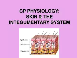
Physiology of Skin Grafts. SKIN: Physiology & Function. Epidermis: protective barrier (against mechanical damage, microbe invasion, & water loss) high regenerative capacity Producer of skin appendages (hair, nails, sweat & sebaceous glands). SKIN: Physiology & Function. Dermis:

E N D
SKIN: Physiology & Function • Epidermis: • protective barrier (against mechanical damage, microbe invasion, & water loss) • high regenerative capacity • Producer of skin appendages (hair, nails, sweat & sebaceous glands)
SKIN: Physiology & Function • Dermis: • mechanical strength (collagen & elastin) • Barrier to microbe invasion • Sensation (point, temp, pressure, proprioception) • Thermoregulation (vasomotor activity of blood vessels and sweat gland activity)
SKIN: Physiology & Function • Immunological surveillance • Most skin is thin, hair-bearing, has sebaceous glands • Skin of palms/soles/flexor surface of digits is thick, not hair-bearing, no sebaceous glands • Vascular supply confined to dermis
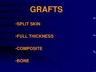
Skin Grafts: Classification • Full thickness skin grafts: - epidermis & full thickness of dermis • Split skin graft: • epidermis & a variable proportion of dermis • thin, intermediate or thick
Skin Grafts: Classification • Autografts • Isografts • Allografts • Xenografts
Skin Grafts: “Process of Take” • Vascularity of donor site • Tolerance to ischaemia • Metabolic activity of the graft
Skin Grafts: “Process of Take” • 4 Phases: • Fibrin adhesion • Plasmatic imbibition • Revascularization: Inosculation & capillary ingrowth • Remodelling: Revascularization & fibrous attachment in restoring normal histological architecture
Skin Grafts: “Process of Take” • Plasmatic Imbibition: • Initially graft ischaemic (24 – 48 hrs) • Fibrin adhesion • Imbibition allows the graft to survive this period • ? Important for nutrition of graft • ? Stops drying out
Skin Grafts: “Process of Take” • Inosculation & capillary ingrowth: • At 48 hrs • Through fibrin layer • Capillary buds from recipient bed contact graft vessels • Open channels (neo-vascularization) pink graft
Skin Grafts: “Process of Take” • Revascularization & fibrous attachment: • Connection of graft & host vessels via anastomoses (inosculation) • Formation of new vascular channels by invasion of graft (neovascularisation) • Combination of old & new vessels (revascularisation) • Fibroblast proliferation: conversion of fibrin adhesion fibrous tissue attachment (anchorage within 4 days)
Skin Graft Take: Dermis • Fibrous component:
Skin Graft Take: Dermis • Appendages: - sweating dependent on no. of transplanted sweat glands & degree of sympathetic reinnervation; will sweat like recipient site in FTSG only - sebaceous gland activity mostly in thicker grafts: SSG usually dry & shiny - hair grows from FTSG if well taken with no complications
Skin Graft Healing • Initially white then pinkens with new blood supply • Lymphatic drainage by day 6 • Collagen replacement from day 7 to week 6 • Vascular remodelling for months
Skin Graft Healing • Contraction: - shrinks immediately due to elastic recoil: – FTSG 40%; medium SSG 20%; thin SSG 10%. - secondary contracture as heals: - FTSG remains same size after above shrinkage; - SSG will contract as much as possible; - more dermis = less contraction - ? Due to myofibroblasts
Skin Graft Healing • Reinnervation: • from margins to bed; • 4/52 to 2 years; • Depends on graft thickness and bed; • Uneventful healing leads to near normal 2PD; • Cold sensitivity can be a problem.
Skin GraftExpansion • Based on principle that wounds reepithelialized from the periphery • Expansion provides larger areas from which epithelium can grow • Larger areas can be covered with less skin
Skin GraftExpansion • Meshing - covers large area - easier to contour - fluid can drain through holes - cosmetic results less than ideal - various mesh ratio
Skin Graft Survival • Meticulous technique • Atraumatic graft handling • Well vascularized bed • Haemostasis • Immobilization • No proximal constricting bandages
Skin Graft Failure • Haematoma • Infection • Seroma • Mobility • Inappropriate bed • Dependency • Arterial insufficiency • Venous congestion • Lymphatic stasis • Technical – upside-down