Download
1 / 105
1.09k likes | 1.33k Vues
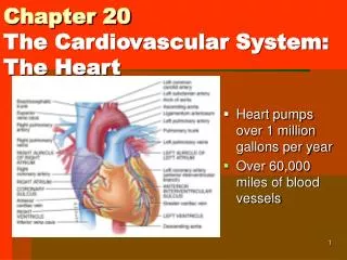
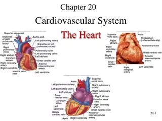
Chapter 20: The Heart Biol 141 A& P R.L. Brashear-Kaulfers. How are the cardiovascular system and heart organized?. Organization of the Cardiovascular System. PLAY. The Heart: Anatomy. Figure 20–1. The Pulmonary Circuit. Carries blood to and from gas exchange surfaces of lungs

E N D
Organization of the Cardiovascular System PLAY The Heart: Anatomy Figure 20–1
The Pulmonary Circuit • Carries blood to and from gas exchange surfaces of lungs The Systemic Circuit • Carries blood to and from the body Alternating Circuits • Blood alternates between pulmonary circuit and systemic circuit
3 Types of Blood Vessels • Arteries: • carry blood away from heart • Veins: • carry blood to heart • Capillaries: • networks between arteries and veins
Capillaries • Also called exchange vessels • Exchange materials between blood and tissues • Dissolved gases, nutrients, wastes
4 Chambers of the Heart • 2 for each circuit: • left and right: ventricles and atria • Right atrium: • collects blood from systemic circuit • Right ventricle: • pumps blood to pulmonary circuit
4 Chambers of the Heart • Left atrium: • collects blood from pulmonary circuit • Left ventricle: • pumps blood to systemic circuit
Anatomy of the Heart • Located directly behind sternum Y PLA Figure 20–2a
Anatomy of the Heart • Great veins and arteries at the base • Pointed tip is apex Figure 20–2c
Relation to Thoracic Cavity Surrounded by pericardial sac Between 2 pleural cavities In the mediastinum Figure 20–2b
The Pericardium • Double lining of the pericardial cavity Figure 20–2c
2 Layers of Pericardium • Parietal pericardium: • outer layer • forms inner layer of pericardial sac • Visceral pericardium: • inner layer of pericardium
Structures of Pericardium • Pericardial cavity: • Is between parietal and visceral layers • contains pericardial fluid • Pericardial sac: • fibrous tissue • surrounds and stabilizes heart Pericarditis An infection of the pericardium
Superficial Anatomy of the Heart • 4 cardiac chambers • Atria - Thin-walled • Expandable outer auricle • Coronary sulcus: • divides atria and ventricles • Anterior and posterior interventricular sulci: • separate left and right ventricles • contain blood vessels of cardiac muscle Figure 20–3
The Heart Wall Figure 20–4
3 Layers of the Heart Wall • Epicardium:- outer layer • Visceral pericardium , Covers the heart • Myocardium: middle layer, Muscular wall • Concentric layers of cardiac muscle tissue • Atrial myocardium wraps around great vessels • 2 divisions of ventricular myocardium • Endocardium: inner layer
Cardiac Muscle Cells • Intercalated discs: • interconnect cardiac muscle cells • secured by desmosomes • linked by gap junctions • convey force of contraction • propagate action potentials Figure 20–5
Characteristics of Cardiac Muscle Cells • Small size • Single, central nucleus • Branching interconnections between cells • Intercalated discs
Cardiac Cells vs. Skeletal Fibers Table 20-1
What is the path of blood flow through the heart, and what are the major blood vessels, chambers, and heart valves?
Internal Anatomy 3D Panorama of the Heart PLAY Figure 20–6a
Atrioventricular(AV)Valves • Connect right atrium to right ventricle and left atrium to left ventricle • Permit blood flow in 1 direction: • atria to ventricles Septa – • Interatrial septum: • separates atria • Interventricular septum: • separates ventricles
The Vena Cava • Delivers systemic circulation to right atrium • Superior vena cava: • receives blood from head, neck, upper limbs, and chest • Inferior vena cava: • receives blood from trunk, and viscera, lower limbs Coronary Sinus • Cardiac veins return blood to coronary sinus • Coronary sinus opens into right atrium
Foramen Ovale • Before birth, is an opening through interatrial septum • Connects the 2 atria • Seals off at birth, forming fossa ovalis
Cusps - Fibrous flaps that form bicuspid (2) and tricuspid (3) valves- Prevent valve from opening backward Right Atrioventricular(AV)Valve • Also called tricuspid valve • Opening from right atrium to right ventricle • Has 3 cusps • Prevents backflow
The Pulmonary Circuit • Conus arteriosus (superior right ventricle) leads to pulmonary trunk • Pulmonary trunk divides into left and right pulmonary arteries • Blood flows from right ventricle to pulmonary trunk through pulmonary valve • Pulmonary valve has 3 semilunar cusps
Return from Pulmonary Circuit • Blood gathers into left and right pulmonary veins • Pulmonary veins deliver to left atrium • Blood from left atrium passes to left ventricle through left atrioventricular(AV)valve • 2-cusp bicuspid valve or mitral valve
The Left Ventricle • Holds same volume as right ventricle • Is larger; muscle is thicker, and more powerful • Similar internally to right ventricle, but does not have moderator band Systemic circulation: • blood leaves left ventricle through aortic valve into ascending aorta • ascending aorta turns (aortic arch) and becomes descending aorta
Left and Right Ventricles • Have significant structural differences • Right ventricle wall is thinner, develops less pressure than left ventricle • Right ventricle is pouch-shaped, left ventricle is round Figure 20–7
The Heart Valves • One-way valves prevent backflow during contraction • (AV)Valves- between atria and ventricles • Blood pressure closes valve cusps during ventricular contraction • Papillary muscles tense chordae tendineae: • prevent valves from swinging into atria • Regurgitation -Failure of valves • Causes backflow of blood into atria Figure 20–8
Semilunar Valves • Pulmonary and aortic tricuspid valves • Prevent backflow from pulmonary trunk and aorta into ventricles • Have no muscular support • 3 cusps support like tripod
Aortic Sinuses - at base of ascending aorta • Prevent valve cusps from sticking to aorta • Origin of right and left coronary arteries Carditis - An inflammation of the heart • Can result in valvular heart disease(VHD): e.g.,rheumatic fever
KEY CONCEPT • The heart has 4 chambers: • 2 for pulmonary circuit: • right atrium and right ventricle • 2 for systemic circuit: • left atrium and left ventricle • Left ventricle has a greater workload • Is much more massive than right ventricle, but the two chambers pump equal amounts of blood • AV valves prevent backflow from ventricles into atria • Semilunar valves prevent backflow from aortic and pulmonary trunks into ventricles
Connective Tissue Fibers of the Heart • Physically support cardiac muscle fibers • Distribute forces of contraction • Add strength and prevent overexpansion of heart • Elastic fibers return heart to original shape after contraction
Blood Supply to the Heart • Coronary circulation Figure 20–9
Coronary Circulation • Coronary arteries-Left and right Originate at aortic sinuses • High blood pressure, elastic rebound force blood through coronary arteries between contractions • cardiac veins • Supplies blood to muscle tissue of heart
Right Coronary Artery • Supplies blood to: • right atrium • portions of both ventricles • cells of sinoatrial(SA) and atrioventricular nodes • marginal arteries (surface of right ventricle) • posterior interventricular artery
Left Coronary Artery • Supplies blood to: • left ventricle • left atrium • interventricular septum • 2 main branches: • circumflex artery • anterior interventricular artery
Cardiac Veins • Great cardiac vein: • drains blood from area of anterior interventricular artery into coronary sinus • Anterior cardiac vein: • empties into right atrium • Posterior cardiac vein, middle cardiac vein, and small cardiac vein: • empty into great cardiac vein or coronary sinus
The Cardiac Cycle • The Heartbeat • A single contraction of the heart • The entire heart contracts in series: • first the atria • then the ventricles Figure 20–11
2 Types of Cardiac Muscle Cells • Conducting system: • controls and coordinates heartbeat • Contractile cells: • produce contractions * The Cardiac Cycle begins with action potential at SA node • transmitted through conducting system • produces action potentials in cardiac muscle cells (contractile cells) • Electrical events in the cardiac cycle can be recorded on an electrocardiogram(ECG)
The Conducting System Figure 20–12
The Conducting System • A system of specialized cardiac muscle cells: • initiates and distributes electrical impulses that stimulate contraction • Automaticity: • cardiac muscle tissue contracts automatically
Structures of the Conducting System • Sinoatrial (SA) node • Atrioventricular (AV) node • Conducting cells
Conducting Cells • Interconnect SA and AV nodes • Distribute stimulus through myocardium • In the atrium: • internodal pathways • In the ventricles: • AV bundle and bundle branches
Prepotential • Also called pacemaker potential • Resting potential of conducting cells: • gradually depolarizes toward threshold • SA node depolarizes first, establishing heart rate
Heart Rate • SA node generates 80–100 action potentials per minute • Parasympathetic stimulation slows heart rate • AV node generates 40–60 action potentials per minute
Impulse Conduction through the Heart Figure 20–13
The Sinoatrial(SA)Node • In posterior wall of right atrium • Contains pacemaker cells • Connected to AV node by internodal pathways • Begins atrial activation (Step 1)