Download
1 / 44
440 likes | 514 Vues
Circulation and gas transport (continued). IB-202-14 3/19/06. 3. The clotting process begins when the endothelium of a vessel is damaged, exposing connective tissue in the vessel wall to blood. Platelets adhere to collagen fibers in the connective tissue and release a substance that

E N D
Circulation and gas transport (continued) IB-202-14 3/19/06
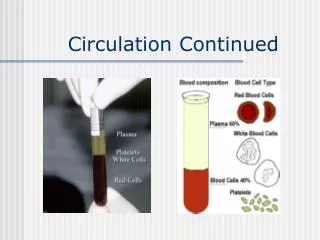
3 The clotting process begins when the endothelium of a vessel is damaged, exposing connective tissue in the vessel wall to blood. Platelets adhere to collagen fibers in the connective tissue and release a substance that makes nearby platelets sticky. The platelets form a plug that provides emergency protection against blood loss. This seal is reinforced by a clot of fibrin when vessel damage is severe. Fibrin is formed via amultistep process: Clotting factors released fromthe clumped platelets or damaged cells mix withclotting factors in the plasma, forming an activation cascade that converts a plasma proteincalled prothrombin to its active form, thrombin.Thrombin itself is an enzyme that catalyzes the final step of the clotting process, the conversion of fibrinogen to fibrin. The threads of fibrin become interwoven into a patch (see colorized SEM). 2 1 Collagen fibers Fibrin clot Plateletplug Red blood cell Platelet releases chemicalsthat make nearby platelets sticky Clotting factors from: Platelets Damaged cells Plasma (factors include calcium, vitamin K) Prothrombin Thrombin Figure 42.17 Fibrin Fibrinogen 5 µm Blood Clotting • A cascade of complex reactions • Converts fibrinogen to fibrin, forming a clot A baby aspirin per day makes the platelets lazy! Hemophiliacs
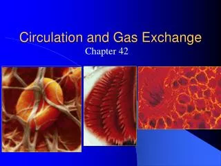
Cardiovascular Disease • Cardiovascular diseases • Are disorders of the heart and the blood vessel and account for more than half the deaths in the United States
Smooth muscle Connective tissue Plaque Endothelium (a) Normal artery (b) Partly clogged artery 50 µm 250 µm Figure 42.18a, b • One type of cardiovascular disease, atherosclerosis • Is caused by the buildup of cholesterol within arteries (low density lipoprotein complexes with cholesterol) Plaques sites of inflammation and can cause a clot to form if plaque splits open! Aspirin! Thrombus!
Hypertension, or high blood pressure • Promotes plaque formation and increases the risk of heart attack and stroke • A heart attack • Is the death of cardiac muscle tissue resulting from blockage of one or more coronary arteries • Either by plaque build up or a clot (thrombus) formed elsewhere and lodging in the vessel. Angina (pain in chest) Nitroglycerin-explosive! Releases nitric oxide relaxe arterioles. • A stroke • Is the death of nervous tissue in the brain, usually resulting from rupture or blockage of arteries in the head (clot dissolving enzymes useful if administered immediately).
Respiratorymedium(air of water) O2 CO2 Respiratorysurface Organismal level Circulatory system Cellular level Energy-richmoleculesfrom food ATP Cellular respiration Figure 42.19 • Concept 42.5: Gas exchange occurs across specialized respiratory surfaces • Gas exchange • Supplies oxygen for cellular respiration and disposes of carbon dioxide Oxygen is final electron acceptor in electron transport chain!!
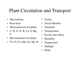
Animals require large, moist respiratory surfaces for the adequate diffusion of respiratory gases • Between their cells and the respiratory medium which can be either air or water • Gills are outfoldings of the body surface specialized for gas exchange in aquatic animals
(a) Sea star. The gills of a sea star are simple tubular projections of the skin. The hollow core of each gillis an extension of the coelom(body cavity). Gas exchangeoccurs by diffusion across thegill surfaces, and fluid in thecoelom circulates in and out ofthe gills, aiding gas transport. The surfaces of a sea star’s tube feet also function in gas exchange. Gills Coelom Figure 42.20a Tube foot • In some invertebrates • The gills have a simple shape and are distributed over much of the body
(b) Marine worm. Many polychaetes (marine worms of the phylum Annelida) have a pair of flattened appendages called parapodia on each body segment. The parapodia serve as gillsand also function incrawling and swimming. Parapodia Figure 42.20b Gill • Many segmented marine worms (Annelids) have flaplike gills • That extend from each segment of their body
(c) Scallop. The gills of a scallop are long, flattened plates that project from the main body mass inside the hard shell. Cilia on the gills circulate water around the gill surfaces. (d) Crayfish. Crayfish and other crustaceans have long, feathery gills covered by the exoskeleton. Specialized body appendages drive water over the gill surfaces. Gills Gills Figure 42.20c, d • The gills of clams, crayfish, and many other animals • Are restricted to a local body region
Oxygen-poorblood Gill arch Oxygen-richblood Lamella Blood vessel Gill arch 15% 40% 70% 5% Water flow 30% Operculum 100% 60% 90% O2 Blood flowthrough capillariesin lamellaeshowing % O2 Water flowover lamellaeshowing % O2 Gillfilaments Figure 42.21 Countercurrent exchange. Very efficient for extracting oxygen from water! • The effectiveness of gas exchange in some gills, including those of fishes • Is increased by ventilation and countercurrent flow of blood and water Ram jet ventilation!
Air sacs Tracheae Spiracle (a) The respiratory system of an insect consists of branched internal tubes that deliver air directly to body cells. Rings of chitin reinforce the largest tubes, called tracheae, keeping them from collapsing. Enlarged portions of tracheae form air sacs near organs that require a large supply of oxygen. Air enters the tracheae through openings called spiracles on the insect’s body surface and passes into smaller tubes called tracheoles. The tracheoles are closed and contain fluid (blue-gray). When the animal is active and is using more O2, most of the fluid is withdrawn into the body. This increases the surface area of air in contact with cells. Figure 42.22a Tracheal Systems in Insects • The tracheal system of insects • Consists of tiny branching tubes that penetrate the body
Body cell Airsac Tracheole Body wall Myofibrils Trachea Air Tracheoles Mitochondria (b) This micrograph shows cross sections of tracheoles in a tiny piece of insect flight muscle (TEM). Each of the numerous mitochondria in the muscle cells lies within about 5 µm of a tracheole. Figure 42.22b 2.5 µm • The tracheal tubes • Supply O2 directly to body cells
Lungs • Spiders, land snails, and most terrestrial vertebrates have internal lungs.
Branch from thepulmonaryartery(oxygen-poor blood) Branch from the pulmonary vein (oxygen-rich blood) Terminal bronchiole Nasalcavity Alveoli Pharynx Left lung Esophagus Larynx 50 µm Trachea 50 µm Right lung Bronchus Bronchiole Colorized SEM SEM Diaphragm Heart Figure 42.23 Spiders, land snails, and most terrestrial vertebrates have internal lungs. In mammals a system of branching ducts conveys air to the lungs Capillary web over alveoli
In mammals, air inhaled through the nostrils • Passes through the pharynx into the trachea, bronchi, bronchioles, and dead-end alveoli, where gas exchange occurs across a thin layer of water and the plasma membrane!
How an Amphibian Breathes • An amphibian such as a frog • Ventilates its lungs by positive pressure breathing, which forces air down the trachea. • Mammals ventilate using negative pressure breathing. • Reptiles also use negative pressure but have no diaphragm
Rib cage expands asrib muscles contract Rib cage gets smaller asrib muscles relax Air inhaled Air exhaled Lung Diaphragm INHALATIONDiaphragm contracts(moves down) EXHALATIONDiaphragm relaxes(moves up) Figure 42.24 How a Mammal Breathes • Mammals ventilate their lungs • By negative pressure breathing, which pulls air into the lungs
Air Air Anteriorair sacs Trachea Lungs Lungs Posteriorair sacs Air tubes(parabronchi)in lung 1 mm EXHALATIONAir sacs empty; lungs fill INHALATIONAir sacs fill Figure 42.25 How a Bird Breathes • Besides lungs, bird have eight or nine air sacs • That function as bellows that keep air flowing through the lungs in a one way direction
Air passes through the lungs • In one direction only • Every exhalation • Completely renews the air in the lungs. • Thus birds are much more efficient in extracting oxygen from the air and thus can fly at altitudes of 30,000 feet. Humans can barely climb stair at this elevation (Mount Everest climbers need oxygen!)
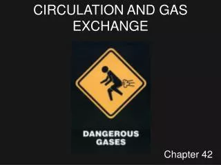
Cerebrospinalfluid The medulla’s control center also helps regulate blood CO2 level. Sensorsin the medulla detect changes in the pH (reflecting CO2 concentration) of the blood and cerebrospinal fluid bathing the surface of the brain. 1 The control center in the medulla sets the basicrhythm, and a control centerin the pons moderates it,smoothing out thetransitions between inhalations and exhalations. 5 Nerve impulses relay changes in CO2 and O2 concentrations. Other sensors in the walls of the aortaand carotid arteries in the neck detect changes in blood pH andsend nerve impulses to the medulla. In response, the medulla’s breathingcontrol center alters the rate anddepth of breathing, increasing bothto dispose of excess CO2 or decreasingboth if CO2 levels are depressed. Pons Breathing control centers Nerve impulses trigger muscle contraction. Nervesfrom a breathing control centerin the medulla oblongata of the brain send impulses to thediaphragm and rib muscles, stimulating them to contractand causing inhalation. 2 Medullaoblongata Carotidarteries Aorta In a person at rest, these nerve impulses result in about 10 to 14 inhalationsper minute. Between inhalations, the musclesrelax and the person exhales. 3 The sensors in the aorta andcarotid arteries also detect changesin O2 levels in the blood and signal the medulla to increase the breathing rate when levels become very low. 6 Figure 42.26 Diaphragm Rib muscles Control of Breathing in Humans • The main breathing control centers • Are located in two regions of the brain, the medulla oblongata and the pons 4
The centers in the medulla • Regulate the rate and depth of breathing in response to pH changes in the cerebrospinal fluid • The medulla adjusts breathing rate and depth • To match metabolic demands
Sensors in the aorta and carotid arteries • Monitor O2 and CO2 concentrations in the blood • Exert secondary control over breathing • At high altitudes the oxygen sensors kick in and causes deep rapid breathing. This “blows” off excess carbon dioxide making ones blood alkaline and gives one head aches (don’t feel well either). Some people more susceptible to altitude sickness than others (10,000 ft for some).
Respiratory pigments bind and transport gases • The metabolic demands of many organisms require that the blood transport large quantities of O2 and CO2. The amount is more than can be physically dissolved in solution! Thus the need for respiratory pigments.
Composition of air and solubility of gases in water • Air pressure at sea level =760 mmHg • Air is 78% Nitrogen, 21% Oxygen and .2% Carbon dioxide. Each gas exerts it pressure independently of the other. • Thus the partial pressure of O2 =.21 X 760= ~160 mmHg, N2 600 mmHg and CO2 .23mmHg • Solubility of pure gas in one liter of water. O2 = 49 ml/l, N2 =24 ml/l,CO2 = 1713 ml/l • Temperature decreases, increase solubility • Salt decreases solubility • Thus, an aquatic animal living in a tropical tide pool doesn’t have access to much oxygen in the water.
A gas always diffuses from a region of higher partial pressure too a region of lower partial pressure • Gases diffuse down pressure gradients in the lungs and other organs
In the lungs and in the tissues • O2 and CO2 diffuse from where their partial pressures are higher to where they are lower
Exhaled air contains a lot of oxygen because of mixing in dead end space! Inhaled air Exhaled air 120 27 160 0.2 Alveolar spaces O2 CO2 O2 CO2 Alveolarepithelialcells 104 40 O2 CO2 O2 CO2 Blood leaving alveolar capillaries Blood enteringalveolarcapillaries O2 CO2 3 4 2 1 Alveolar capillariesof lung 40 45 104 40 O2 O2 CO2 CO2 Pulmonaryveins Pulmonaryarteries Systemic arteries Systemicveins Heart Tissue capillaries O2 CO2 Blood enteringtissuecapillaries Blood leavingtissuecapillaries O2 CO2 100 40 40 45 O2 O2 CO2 CO2 Tissue cells <40 >45 O2 CO2 0.5 liter tidal volume 4.8 l vital capacity 1.2 l residual space Figure 42.27
Need for Respiratory Pigments • Respiratory pigments are proteins that bind and transport oxygen • Can only dissolve 4.5 ml of oxygen in a liter of blood without Hb. • Greatly increase the amount of oxygen that blood can carry (200 ml of oxygen /liter)
Oxygen Transport • The respiratory pigment of almost all vertebrates is the protein hemoglobin, contained in the erythrocytes • In invertebrates that have pigments they are hemocyanin, a large copper containing protein that circulates free in solution ( not housed in cells like hemoglobin).
Heme group Iron atom O2 loaded in lungs O2 O2 unloaded In tissues O2 Polypeptide chain Hemoglobin a tetrameric molecule • Like all respiratory pigments • Hemoglobin must reversibly bind O2, loading O2 in the lungs and unloading it in other parts of the body Figure 42.28
Loading and unloading of O2 • Depend on cooperation between the subunits of the hemoglobin molecule • The binding of O2 to one subunit induces the other subunits to bind O2 with more affinity
Cooperative O2 binding and release • Is evident in the dissociation curve for hemoglobin • A drop in pH • Lowers the affinity of hemoglobin for O2
(a) PO2 and Hemoglobin Dissociation at 37°C and pH 7.4 O2 unloaded from hemoglobin during normal metabolism 100 80 O2 reserve that can be unloaded from hemoglobin to tissues with high metabolism 60 O2 saturation of hemoglobin (%) 40 20 0 60 100 40 80 0 20 Tissues at rest Lungs Tissues during exercise PO2 (mm Hg) (b) pH and Hemoglobin Dissociation 100 pH 7.4 80 Bohr shift:Additional O2released from hemoglobin at lower pH(higher CO2concentration) 60 O2 saturation of hemoglobin (%) pH 7.2 40 20 0 Figure 42.29a, b 60 100 40 80 0 20 PO2 (mm Hg)
Arterial Blood O2 saturation and O2 content 20 100 O 2 80 Content 15 Arterial (vol. %) (= mL O2 per 100mL blood) blood 60 10 % O2 saturation 40 5 20 0 0 30 60 90 PO2 (mm Hg)
Carbon Dioxide Transport • Hemoglobin also helps transport CO2 and assists in buffering the blood by forming bicarbonate.
Carbon from respiring cells • Diffuses into the blood plasma and then into erythrocytes and is ultimately released in the lungs
Tissue cell Carbon dioxide produced bybody tissues diffuses into the interstitial fluid and the plasma. Most of the HCO3– diffuseinto the plasma where it is carried in the bloodstream to the lungs. CO2 transportfrom tissues 11 10 7 6 5 4 3 2 1 8 9 CO2 produced Interstitialfluid CO2 Over 90% of the CO2 diffuses into red blood cells, leaving only 7%in the plasma as dissolved CO2. Blood plasmawithin capillary CO2 Capillarywall In the lungs HCO3– diffusesfrom the plasma into red blood cells, combining with H+ released from hemoglobin and forming H2CO3. CO2 H2O Some CO2 is picked up and transported by hemoglobin. Redbloodcell Hemoglobinpicks upCO2 and H+ H2CO3 Hb Carbonic acid Carbonic acid is converted back into CO2 and water. HCO3– + H+ Bicarbonate However, most CO2 reacts with water in red blood cells, forming carbonic acid (H2CO3), a reaction catalyzed bycarbonic anhydrase contained. Withinred blood cells. HCO3– To lungs CO2 formed from H2CO3 is unloadedfrom hemoglobin and diffuses into the interstitial fluid. CO2 transportto lungs HCO3– 9 6 2 7 5 4 3 1 8 + H+ HCO3– CO2 diffuses into the alveolarspace, from which it is expelledduring exhalation. The reductionof CO2 concentration in the plasmadrives the breakdown of H2CO3 Into CO2 and water in the red bloodcells (see step 9), a reversal of the reaction that occurs in the tissues (see step 4). Carbonic acid dissociates into a biocarbonate ion (HCO3–) and a hydrogen ion (H+). HemoglobinreleasesCO2 and H+ Hb H2CO3 H2O CO2 Hemoglobin binds most of the H+ from H2CO3 preventing the H+from acidifying the blood and thuspreventing the Bohr shift. CO2 11 10 CO2 CO2 Figure 42.30 Alveolar space in lung
Elite Animal Athletes • Migratory and diving mammals • Have evolutionary adaptations that allow them to perform extraordinary feats. • Weddell seals dive to 600 meters repeatedly during a 20 minute dive and rest and breath for only about 10 minutes. There is no anaerobic metabolism during this dive pattern. • If a very long dive then they can utilize anaerobic metabolism which provides energy, but then they need to rest for hours to get rid of lactic acid build up.
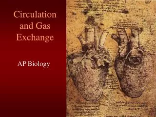
Adaptations for deep diving • Stores twice as much oxygen as humans—in greater volume of RBCs (twice as much blood as a human) and myoglobin in the muscles. RBCs also stored in spleen. • During dive the heart rate slows to 10 beats /min and blood flow is maintained to brain, eyes, spinal cord, adrenal glands and placenta if pregnant • How does the mother ensure that the fetus gets an adequate supply of oxygen during a dive?
100 Fetus 80 Mother 60 O2 saturation of hemoglobin (%) 40 20 0 20 40 60 0 80 100 PO2(mm Hg) Dissociation curve for Weddell seal blood of fetus and mother. Fetus hemoglobin has a greater Bohr effect.
The Ultimate Endurance Runner • The extreme O2 consumption of the antelope-like pronghorn underlies its ability to run at high speed (60km/hr) over long distances. Greater lung surface, higher cardiac output and more mitochrondia per unit muscle. Figure 42.31