Download
1 / 40
400 likes | 595 Vues
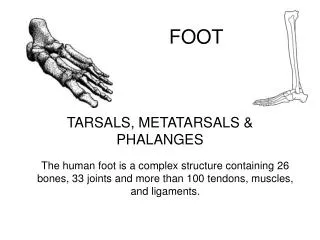
My foot hurts…. Heather Patterson PGY-2 Emergency Medicine May 31, 2007. Objectives. Review relevant foot boney anatomy Brief discussion about 3 foot fractures Practise!. Anatomy. Anatomy. Case. 35M working on roof, falls, lands like a cat c/o bilat heel pain and back pain.

E N D
My foot hurts…. Heather Patterson PGY-2 Emergency Medicine May 31, 2007
Objectives • Review relevant foot boney anatomy • Brief discussion about 3 foot fractures • Practise!
Case • 35M working on roof, falls, lands like a cat • c/o bilat heel pain and back pain
Calcaneus fractures Posterior tuberosity apex of anterior process apex of posterior facet
Calcaneus Fracture • Mechanism: • High energy axial load • Intra or extraarticular • Associations: • 7% bilateral • 10% spine compression # • 25% other LE injury
Calcaneus Fracture • Imaging: • Standard AP/Lat foot and ankle views • Axial • +/- CT • Important distinctions: • Involvement of subtalar joint • Depression of posterior facet
Calcaneus Fracture • Ortho: • Treatment patterns vary • Intraarticular and comminuted fractures must be seen • Outcomes: • Poor outcomes • >50% have loss of ROM, chronic pain, and functional disability
Case • 32M fell and landed with pointed toes
Talar fractures • Anatomy: • 7 articular surfaces (60% of surface) • Regions: • Body • Neck • Head
Talar fractures • Minor talar fractures: • HEAD AND NECK: • Avulsion and chip fractures of superior surface • BODY: • Lateral, medial, posterior body AND osteochondral of talar dome • Require immobilization and referral to ortho for f/u
Talar fractures • Talar neck fractures • 50% of major talar injuries. • Mechanism: • extreme dorsiflexion • Hawkins classification • Often associated fractures
Talar fractures • Type 1: nondisplaced • Type 2: subtalar subluxation • Type 3: dislocation of the talar body (50% open #’s) • Type 4: dislocation of the talar body & distraction of the • talonavicular joint. • Fracture type influences management & prognosis
Talar fractures • Talar body fractures • 23% of all talar fractures • Ie posterior or lateral process fracture • Major talar body fractures are uncommon • usually axial loading
Talar fractures • Talar head fractures • Uncommon (5-10%) • Compression transmitted through the talonavicular joint applied on a plantarflexed foot
Talar fractures • Management: • Major fractures require ortho consult • Outcomes: • Risk of AVN, OA, and chronic pain
Case • 18F playing soccer, tripped and twisted foot • Not sure of how she twisted/landed
Navicular Fracture • Classification: • Dorsal avulsion • >50% of navicular #s • Eversion injury • Associated with deltoid ligament injury • Minimal articular involvement • Tuberosity Fracture • Eversion injury • Associated with posterior tibialis tendon avulsion
Navicular Fracture • Classification: • Body Fracture • Rare • Axial loading • Comminuted, intraarticular
Navicular Fracture • Clinical • Pain on palpation • +/- pain on passive eversion or active inversion • Imaging • Standard foot views • +/- bone scan
Navicular Fracture • Why do we care? • Significant risk of AVN • Management: • Outpatient Ortho: • Dorsal avulsion and tuberosity # with minimal articular involvement • Immobilize 4-6 wks • ED Ortho consult • Body#, displaced #, >20% of articular surface involved