Download
1 / 2
20 likes | 167 Vues
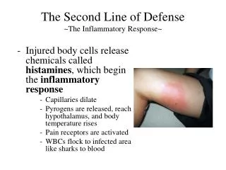
BEAS-2B. Cultured in 0, 1, 2.5 or 5 mM GLN with 1 μg LPS/mL. DMEM/F12. 14hr. 12hr. 16hr. IL-8. 0 mM GLN. β -actin. 1 mM GLN. 5 mM GLN. Effects of Glutamine on the Inflammatory Response of Pulmonary Epithelial Cells. Yu-Chen Hou and Sung-Ling Yeh

E N D
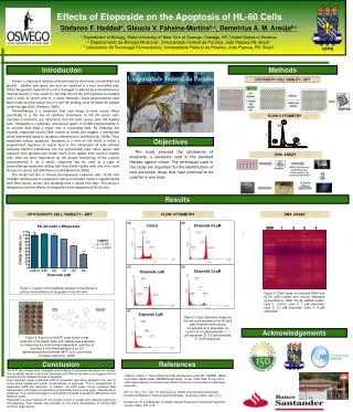
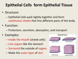
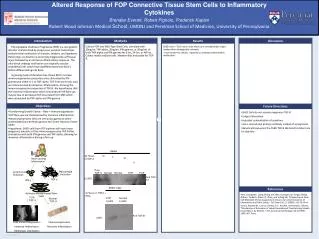
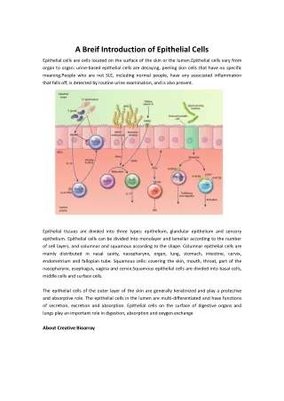
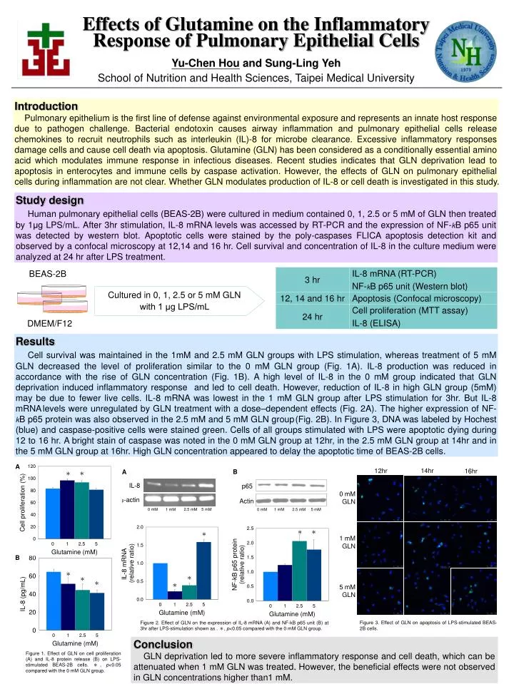
BEAS-2B Cultured in 0, 1, 2.5 or 5 mM GLN with 1 μg LPS/mL DMEM/F12 14hr 12hr 16hr IL-8 0 mM GLN β-actin 1 mM GLN 5 mM GLN Effects of Glutamine on the Inflammatory Response of Pulmonary Epithelial Cells Yu-Chen Hou and Sung-Ling Yeh School of Nutrition and Health Sciences, Taipei Medical University Introduction Pulmonary epithelium is the first line of defense against environmental exposure and represents an innate host response due to pathogen challenge. Bacterial endotoxin causes airway inflammation and pulmonary epithelial cells release chemokines to recruit neutrophils such as interleukin (IL)-8 for microbe clearance. Excessive inflammatory responses damage cells and cause cell death via apoptosis. Glutamine (GLN) has been considered as a conditionally essential amino acid which modulates immune response in infectious diseases. Recent studies indicates that GLN deprivation lead to apoptosis in enterocytes and immune cells by caspase activation. However, the effects of GLN on pulmonary epithelial cells during inflammation are not clear. Whether GLN modulates production of IL-8 or cell death is investigated in this study. Study design Human pulmonary epithelial cells (BEAS-2B) were cultured in medium contained 0, 1, 2.5 or 5 mM of GLN then treated by 1μg LPS/mL. After 3hr stimulation, IL-8 mRNA levels was accessed by RT-PCR and the expression of NF-kB p65 unit was detected by western blot. Apoptotic cells were stained by the poly-caspases FLICA apoptosis detection kit and observed by a confocal microscopy at 12,14 and 16 hr. Cell survival and concentration of IL-8 in the culture medium were analyzed at 24 hr after LPS treatment. Results Cell survival was maintained in the 1mM and 2.5 mM GLN groups with LPS stimulation, whereas treatment of 5 mM GLN decreased the level of proliferation similar to the 0 mM GLN group (Fig. 1A). IL-8 production was reduced in accordance with the rise of GLN concentration (Fig. 1B). A high level of IL-8 in the 0 mM group indicated that GLN deprivation induced inflammatory response and led to cell death. However, reduction of IL-8 in high GLN group (5mM) may be due to fewer live cells. IL-8 mRNA was lowest in the 1 mM GLN group after LPS stimulation for 3hr. But IL-8 mRNAlevels were unregulated by GLN treatment with a dose–dependent effects (Fig. 2A). The higher expression of NF-kB p65 protein was also observed in the 2.5 mM and 5 mM GLN group(Fig. 2B). In Figure 3, DNA was labeled by Hochest (blue) and caspase-positive cells were stained green. Cells of all groups stimulated with LPS were apoptotic dying during 12 to 16 hr. A bright stain of caspase was noted in the 0 mM GLN group at 12hr, in the 2.5 mM GLN group at 14hr and in the 5 mM GLN group at 16hr. High GLN concentration appeared to delay the apoptotic time of BEAS-2B cells. A B A * * p65 Actin 0 mM 5 mM 2.5 mM 1 mM 0 mM 5 mM 2.5 mM 1 mM * * * B * * * * * Figure 3. Effect of GLN on apoptosis of LPS-stimulated BEAS-2B cells. Figure 2. Effect of GLN on the expression of IL-8 mRNA (A) and NF-kB p65 unit (B) at 3hr after LPS-stimulation shown as . *, p<0.05 compared with the 0 mM GLN group. Conclusion GLN deprivation led to more severe inflammatory response and cell death, which can be attenuated when 1 mM GLN was treated. However, the beneficial effects were not observed in GLN concentrations higher than1 mM. Figure 1. Effect of GLN on cell proliferation (A) and IL-8 protein release (B) on LPS-stimulated BEAS-2B cells. *, p<0.05 compared with the 0 mM GLN group.