Download

1 / 12
120 likes | 256 Vues
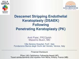
Comparison of Donor Insertion Techniques on Endothelial Cell Mortality in DSAEK. Authors Abraham Sleem , MD Robert L. Schultze , MD Stephen S. Khachikian , MD William Yang, MD. No Financial Interest in Study Material. Many Insertion Techniques & Devices.

E N D
Comparison of Donor Insertion Techniques on Endothelial Cell Mortality in DSAEK Authors Abraham Sleem, MD Robert L. Schultze, MD Stephen S. Khachikian, MD William Yang, MD No Financial Interest in Study Material
Many Insertion Techniques & Devices • Purpose: To determine the impact of donor insertion technique on post-operative endothelial cell density in patients undergoing DSAEK. • Methods used: • Ogawa Forceps/Taco • Busin Glide (Moria) • Others Include: • Suture Pull Through • Neusidl Corneal Inserter (Fischer)
Factors Thought to Influence Endothelial Cell Survival • Recent work by Terry et al suggest larger surgical wound (5mm vs 3mm) is associated with less endothelial cell loss • Precut tissue does not show a difference in cell survival • Does the reduced trauma (no folding, less crushing) to the donor tissue by the Busin glide technique induce less insertion trauma to donor? Busin M, Bhatt PR, Scorcia V. A modified technique for descemet membrane stripping automated endothelial keratoplasty to minimize endothelial cell loss. Arch Ophthalmol. 2008:1133-7. Mehta JS, Por YM, Poh R, Beuerman RW, Tan D. Comparison of donor insertion techniques for descemet stripping automated endothelial keratoplasty. Arch Ophthalmol. 2008:1383-8 Terry MA, Saad HA, Shamie N, Chen ES, Phillips PM, Friend DJ, Holiman JD, Stoeger C. Endothelial keratoplasty: the influence of insertion techniques and incision size on donor endothelial survival. Cornea. 2009:24-31. Terry MA, Chen ES, Shamie N, Hoar KL, Friend DJ. Endothelial cell loss after Descemet's stripping endothelial keratoplasty in a large prospective series. Ophthalmology. 2008:488-496. Chen E, Terry M, Shamie N, Hoar K, Phillips P, Friend D. Endothelial Keratoplasty: Vision, Endothelial Survival, and Complications in a Comparative Case Series of Fellows vs Attending Surgeons. American Journal of Ophthalmology. 2009: VOL. XX, NO. X Bahar I, Kaiserman I, Sansanayudh W, Levinfger E, Rootman D. Busin Guide vs Forceps for the Insertion of the Donor Lenticule in Descemet Stripping Automated Endothelial Keratoplasty. American Journal of Ophthalmology. 2009:220-228.
Methods • 45 consecutive eyes underwent DSAEK by one surgeon (RLS) • 24 patients received a donor button folded 60/40 utilizing non-opposing “Ogawa” forceps (Moria) • The remaining 21 patients had the donor inserted utilizing a “rolled, pull-through technique” with a Busin Glide and forceps (Moria)
Methods • All donor tissue came from the Lions Eye Bank in Albany, NY • Pre-operative donor endothelial cell density was determined at the procuring the Eye Bank and post-operative measurements were determined in office 6 months postoperatively. • SP4000 noncontact specular microscope, Konan Medical Corp.
Surgical Technique • DSAEK donor buttons prepared with 300 head on Moria ALTK system • All Donors punched to 8mm • Donor buttons inserted through 5 mm wound • Cornea massage and venting performed • Air bubble maintained for one hour then burped
Results • 6 month overall mean endothelial cell loss was 34.4±20.9% (preop 3086±249 cells/mm2 and post-operative 2016±646 cells/mm2). • Folded donors demonstrated an average cell loss of 36.5±24.2% • “Rolled/Glide” donors had a 31.9±16.6% cell loss (p=0.46).
Preop vs. Postop BSCVA Snellen Equivalent 20/XX
6 month Cell Loss In Other Studies Busin M, Bhatt PR, Scorcia V. A modified technique for descemet membrane stripping automated endothelial keratoplasty to minimize endothelial cell loss. Arch Ophthalmol. 2008:1133-7. Mehta JS, Por YM, Poh R, Beuerman RW, Tan D. Comparison of donor insertion techniques for descemet stripping automated endothelial keratoplasty. Arch Ophthalmol. 2008:1383-8 Terry MA, Saad HA, Shamie N, Chen ES, Phillips PM, Friend DJ, Holiman JD, Stoeger C. Endothelial keratoplasty: the influence of insertion techniques and incision size on donor endothelial survival. Cornea. 2009:24-31. Terry MA, Chen ES, Shamie N, Hoar KL, Friend DJ. Endothelial cell loss after Descemet's stripping endothelial keratoplasty in a large prospective series. Ophthalmology. 2008:488-496. Chen E, Terry M, Shamie N, Hoar K, Phillips P, Friend D. Endothelial Keratoplasty: Vision, Endothelial Survival, and Complications in a Comparative Case Series of Fellows vs Attending Surgeons. American Journal of Ophthalmology. 2009: VOL. XX, NO. X Bahar I, Kaiserman I, Sansanayudh W, Levinfger E, Rootman D. Busin Guide vs Forceps for the Insertion of the Donor Lenticule in Descemet Stripping Automated Endothelial Keratoplasty. American Journal of Ophthalmology. 2009:220-228.
Conclusions • Although the Glide technique showed less endothelial cell death at 6 months and 1 year, this difference was not statistically significant. • Our cell loss was in agreement with other published studies
Discussion • Subjectively the Glide insertion technique induces less donor trauma, but this did not show a difference in overall endothelial cell loss. • There are likely other factors that act as an “equalizer” in terms of cell loss: • Donor button creation • AC Maintainer Management • Air Bubble Management • Surgeon Experience