Download
1 / 38
380 likes | 529 Vues
Chapter 41 Animal Nutrition. Aisha Cissna Brooke Lusa. Dietary Categories. Herbivores Carnivores Omnivores. Adequate Diet. Fuel Raw organic materials Essential nutrients. Energy Budget- flow of energy. Largest portion of energy production of ATP Mostly from cell respiration

E N D
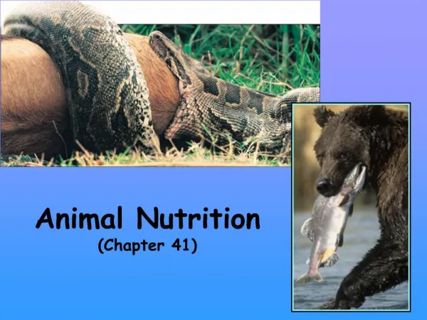
Chapter 41Animal Nutrition Aisha Cissna Brooke Lusa
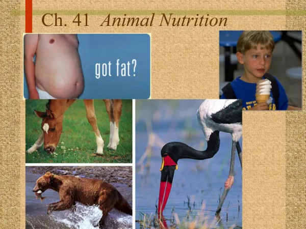
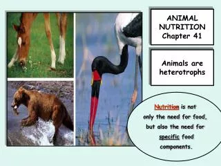
Dietary Categories • Herbivores • Carnivores • Omnivores
Adequate Diet • Fuel • Raw organic materials • Essential nutrients
Energy Budget- flow of energy • Largest portion of energy production of ATP • Mostly from cell respiration • Organic molecules monomers= fuel • Rich organic molecules • Carbs • Fats • Proteins
Homeostasis = the maintenance of stable, internal conditions w/specific limits • GLUCOSE REGULATION • Excess calories used for biosynthesis • Body stores energy in “depots” • Regulated by hormones • When energy needed is more than energy consumed fuel in storage depots is oxidized • Liver glycogen • muscle
2) Insulin enhances the transport of glucose into body cells and stimulates the liver and muscle cells to store glucose as glycogen. As a result, blood glucose level drops. 4) Glucagon promotesthe breakdown ofglycogen in theliver and the release of glucose into the blood, increasing bloodglucose level. Glucose Regulation 1) When blood glucose level rises, a gland called the pancreas secretes insulin, a hormone, into the blood. STIMULUS: Blood glucose level rises after eating. Homeostasis: 90 mg glucose/ 100 mL blood STIMULUS: Blood glucose level drops below set point. 3) When blood glucose level drops the pancreas secretes the hormone glucagon which opposes the effect of insulin.
Caloric Imbalance • Undernourishment- deficient in calories, glycogen and fat used up • Overnourishment- body hoards fat instead of using for fuel
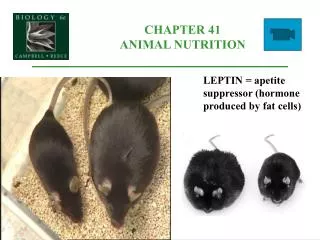
Control? • Feedback circuits control the body’s storage and metabolism of fat • Hormones regulate appetite through “satiety center” in the brain • Most weight regulating hormones= polypeptides (proteins) • Ex: hormone leptin- long term appetite regulator in mammal • Feedback inhibition= produced by adipose = leptin =appetite
41.3 The Main Stages of food processing are ingestion, digestion, absorption, and elimination Ingestion: A heterotrophic mode of nutrition in which other organisms or detritus are eaten whole or in pieces Digestion: The process of breaking down food into molecules small enough for the body to absorb Enzymatic hydrolysis: The process in digestion that splits macromolecules from food by the enzymatic addition of water
Absorption: The uptake of small nutrient molecules by an organism’s own body; the 3rd main stage of food processing following digestion Elimination: The passing of undigested material out of the digestive compartment
Intracellular digestion: The joining of food vacuoles and lysosomes to allow chemical digestion to occur within the cytoplasm of a cell Extracellular digestion: The breakdown of food outside cells.
Gastrovascular cavity: An extensive pouch that serves as the site of extracellular digestion and a passageway to disperse materials throughout most of an animal’s body Complete digestive tract/alimentary canal: A digestive tract consisting of a tube running between a mouth and an anus
We have to break down the macromolecules in our food into monomers. • WHY? • Polymers are too large to pass through cell membranes. • Macromolecules that make up an animal aren’t organized in the same way as those that make up food.
Digestion • Polysaccharides and disaccharides simple sugars • Fats glycerol and fatty acids • Proteins amino acids • Nucleic acids nucleotides • HOW are these broken down? • Enzymatic hydrolysis!
How do animals digest their food without digesting their own cells? • Digestive compartments! -vacuoles • 2 TYPES OF DIGESTION Intracellular digestion-pinocytosis and phagocytosis Extracellular Digestion
Pinocytosis Phagocytosis
Extracellular Digestion • Occurs within compartments continuous with the outside of animal’s body alimentary canal • Mammals have specialized regions to carry out digestion • We can ingest additional food before earlier meals are completely digested (inefficient and difficult for animals w/gastrovascular cavities)
How is the alimentary canal continuous with the outside of the body? • It’s continuous with the outside environment via the mouth and anus, nutrients haven’t entered the body yet by crossing the membrane.
41.4 Each organ of the mammalian digestive system has specialized food-processing functions • Alimentary canal • Peristalsis-Rhythmicwaves of contraction of smooth muscles that push food along digestive tract • Sphincters-ring-like valves consisting of modified muscles in a muscular tube, such as tract; closes off the tube like a draw string • Accessory glands- • 3 pairs of salivary glands • Pancreas= gland w/ dual functions: the nonendocrine portion secretes digestive enzymes and an alkaline solution into the small intestine; the endocrine portion secretes the hormones insulin and glucagon in the blood • Liver=largest organ in the body; produces bile, prepares nitrogenous wastes for disposal, detoxifies poisonous chemicals in the blood • Gallbladder= an organ that stores bile and releases it as it is needed into the small intestine
41.4 Oral Cavity, Pharynx, and Esophagus • Oral cavity: the mouth of an animal • Salivary amylase: a salivary gland enzyme that hydrolyzes starch and glycogen • Bolus: a lubricated ball of chewed food • Pharynx: an area in the vertebrate throat where air and food passages cross • Epiglottis: a cartilaginous flap that blocks the top of the windpipe, the glottis, during swallowing, which prevents the entry of food or fluid into the trachea
Esophagus: a channel that conducts food, by peristalsis, from the pharynx to the stomach Small Intestine: the longest section of the alimentary canal; the principal site of the enzymatic hydrolysis of food molecules and the absorption of nutrients
Duodenum: the 1st section of the small intestine, where acid chyme from the stomach mixes with digestive juices from the pancreas, liver, gallbladder, and gland cells of the intestinal walls • Bile: a mixture of substances that is produced in the liver, stored in the gallbladder, and acts as a detergent to aid in the digestion and absorption of fats
Microvillus: one of many fine, fingerlike projections of the epithelial cells in the lumen (cavity) of the small intestine that increase its surface area • Lacteal: a tiny lymph vessel extending into the core of an intestinal villus and serving as the destination for absorbed chylomicrons • Chylomicrons: one of the small intracellular globules composed of fats are mixed with cholesterol and coated with special proteins
Hepatic portal vein: a large circulatory channel that conveys nutrient-laden blood from the small intestine to the liver, which regulates the blood’s nutrient content
Let the digestion begin! • Epiglottis blocks opening to windpipe so we don’t choke (see motion of Adam’s apple) and food goes down esophagus by peristalsis • Swallowing is at first voluntary, but then involuntary waves of muscle contraction push the bolus into the stomach • Chew-mechanical digestion, increase surface area of food for… • Salivation-chemical digestion begins! • Bolus is formed • Bolus goes down pharynx (we swallow)
Figure 41.16 4) The esophageal sphincter relaxes, allowing bolus down esophagus. 2) The swallowing reflex is triggered when a bolus of food reaches the pharynx. 55) After the food had entered the esophagus, the larynx moves downward and opens the breathing passage. 3) The larynx, the upper part of the respiratory tract, moves upward and tips the epiglottis over the glottis, preventing food from entering the trachea. 1) When a person isn’t swallowing , the esophageal sphincter muscle is contracted, the epiglottis is up, and the glottis is open, allowing air flow through the trachea to the lungs 6) Peristalsis moves the bolus down into stomach.
The Stomach • Stomach- stores food, prepares for digestion, in upper abdomen • Gastric juice- digestive fluid, acidic • Pepsin- enzyme in gastric juice that begins hydrolysis of proteins • Pepsinogen- inactive pepsin first secreted by cells located in gastric pits of stomach • Acid chyme- nutrient rich broth; a mix of recently swallowed food and gastric juice • Pyloric sphincter- opening from stomach to small intestine • Cardiac orifice opening- opening from stomach to esophagus
Stomach Esophagus Cardiac orifice Folds of epithelial tissue Pyloric sphincter 5 µm Small intestine 2 3 1 1 3 2 Interior surface of stomach. The interior surface of the stomach wall is highly folded and dotted with pits leading into tubular gastric glands. Pepsinogen and HCI are secreted into the lumen of the stomach. Epithelium HCl converts pepsinogen to pepsin. Pepsinogen Pepsin (active enzyme) Gastric gland. The gastric glands have three types of cells that secrete different components of the gastric juice: mucus cells, chief cells, and parietal cells. HCl Pepsin then activates more pepsinogen, starting a chain reaction. Pepsin begins the chemical digestion of proteins. Mucus cells secrete mucus, which lubricates and protects the cells lining the stomach. Chief cells secrete pepsino- gen, an inactive form of the digestive enzyme pepsin. Parietal cell Chief cell Figure 41.17 Parietal cells secrete hydrochloric acid (HCl).
Digestion Functions • Secretes gastric juice through epithelium stomach lining • Parietal cells secrete HCL acid-> pepsinogen-> pepsin in lumen *positive feedback inhibition • Many suggestive enzymes secreted in inactive form • Coating of mucus secreted by epithelial cells • Smooth muscles mix stomach contents -> acid chyme
Closed or Open? • Cardiac orifice opens for bolus • Pyloric sphincter helps regulate chyme into the intestine • 2- 6 hrs to empty
The Small Intestine 1) Duodenum A) Pancreatic enzymes include protein-digesting enzymes in inactive form but are activated once they’re in the duodenum (like pepsinogenpepsin) B) Epithelial lining of the duodenum called the brush border secretes digestive enzymes, some which are secreted into the lumen of the duodenum, other digestive enzymes bind to the surface of epithelial cells C) Digestion is completed as peristalsis moves chyme and digestive juices while still in the duodenum
Pancreas Membrane-boundenteropeptidase Inactivetrypsinogen Trypsin Other inactiveproteases Active proteases Lumen of duodenum
Vein carrying blood to hepatic portal vessel Microvilli(brush border) Bloodcapillaries Muscle layers Epithelialcells Largecircularfolds Epithelial cells Lacteal Villi Key Lymph vessel Nutrientabsorption Villi Intestinal wall 2) Absorption A) villi and microvilli increase rate of absorption villi absorb nutrientsmicrovillilactealbloodstream B) nutrients are absorbed across the intestinal epithelium and then the unicellular epithelium of capillaries/lacteal
3) Hormones help control secretion of digestive juices 4) Active and Passive transport of nutrients across epithelial cells A) fructose moves by facilitated diffusion down concentration gradient from lumen of intestine epithelial cells capillaries B) amino acids, peptides, vitamins, glucose are pumped against concentration gradient=intestine can absorb a higher proportion of nutrients Amino acids + sugars epithelium capillaries bloodstream converge to hepatic portal vein liver heart
b) Liver regulates amount of nutrients (i.e. glucose)in blood C) Glycerol+fatty acidsepithelial cellsrecombined into fat w/in epithelial cellsform chylomicronslactealsvessels of lymphatic systemlarge veins that return blood to heart
The Large Intestine • Large intestine/colon- connected to the small intestine at a T shaped junction where a sphincter controls the movement of material; functions mainly in water absorption and formation of feces • Cecum-one arm of the T-pouch • Appendix- cecum’s finger-like extension; contains a mass of white blood cells that contribute to immunity • Feces- wastes of the digestive tract • Rectum- terminal portion of the colon
Function? • Recovers water that has entered the alimentary canal as solvent of digestive juices • Feces becomes solid by peristalsis • Constipation results b/c too much water absorbed by intestine=moves slowly • Feces exit out rectum