Download
1 / 6
60 likes | 290 Vues
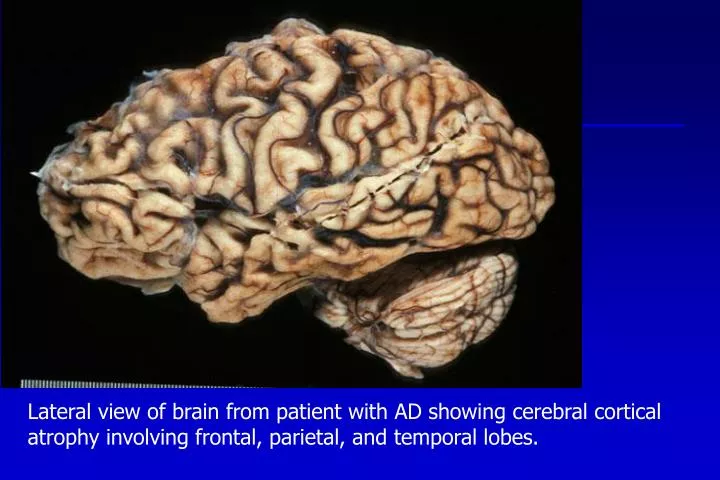
Lateral view of brain from patient with AD showing cerebral cortical atrophy involving frontal, parietal, and temporal lobes. Microscopic image of silver-stained section of cerebrum from a patient with Alzheimer’s disease showing larger neuritic plaque and smaller neurofibrillary tangle.

E N D
Lateral view of brain from patient with AD showing cerebral cortical atrophy involving frontal, parietal, and temporal lobes.
Microscopic image of silver-stained section of cerebrum from a patient with Alzheimer’s disease showing larger neuritic plaque and smaller neurofibrillary tangle.
Microscopic images showing two feature of prion disease: vacuolization (left) and plaque formation (right).
Coronal section of cerebrum at the level of the Anterior commissure showing marked atrophy of heads of caudate nuclei.
PD Control Axial section of midbrain showing loss of pigmentation in the Substantia nigra from a patient who died of Parkinson’s disease; some surviving neurons in this region would harbor Lewy bodies.
Axial whole-mount section of cervical spinal cord (HE/LFB stain) showing pallor of the lateral corticospinal tracts in a patient that died of amyotrophic lateral sclerosis.