Download
1 / 20
200 likes | 372 Vues
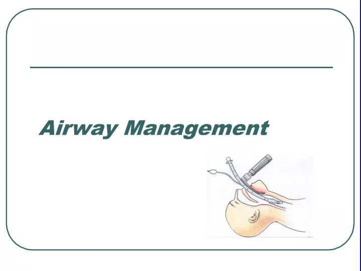
The patient who is difficult to ventilate and difficult to intubate is quite possibly the most serious problem faced by anesthesiologists because hypoxic brain injuries and cardiac arrest are real possibilities in this scenario.

E N D
The patient who is difficult to ventilate and difficult to intubate is quite possibly the most serious problem faced by anesthesiologists because hypoxic brain injuries and cardiac arrest are real possibilities in this scenario. • Although a thorough history and physical examination are likely to identify the majority of patients with difficult airway, unanticipated problems occasionally present.
Only through preplanning and practiced algorithms are such situations managed optimally. • The American Society of Anesthiologists has prepared a difficult airway algorithm to assist the clinician. • Remember !!! This is not the time of heroism, if intubation or ventilation is difficult call for help.
HISTORY : • Any Adverse events related to previous airway management episodes. For instance:- • Have they ever been informed by anesthesiologists that they had an airway management problem( i.e. difficult to intubate, difficult to ventilate ). • Have they had a tracheostomy or other surgery or radiation about the face and neck ? • Have they sustained significant burns to these areas ? • Do they have obstructive sleep apnea or temporomandibular joint ( TMJ ) dysfunction ? • Review of prior anesthetic records is helpful.
Physical Examination:- • 1- Oral Cavity:- • Note extent and symmetry of opening ( three finger-breads is optimal ). • The health of the teeth ( loose, missing, or cracked teeth should be documented ), and the presence of dental appliances. • Prominent buck teeth may interfere with the use of a laryngoscope. • The size of the tongue is noted ( large tongues rarely make airway management impossible, only more difficult ). • High arched palates have been associated with difficulty in visualizing the larynx.
2- The mandible and the quality of TMJ function:- • A short mandibular body ( three finger breads ) as measured from the mental process to the prominence of the thyroid cartilage ( thyromental distance ) suggests difficulty in visualizing the larynx. • Patients with TMJ dysfunction may have asymmetry or limitations in opening the mouth as well as popping and clicking. • Curiously; some patients with TMJ dysfunction have greater difficulty with opening the mouth after anesthetic induction and neuromuscular paralysis than when they are awake and cooperative.
3- Examination of the neck:- • Evidence of prior surgeries ( especially tracheostomy ), or significant burn is noted. • Evidence of any abnormal masses ( e.g. hematoma, abscess or cellulitis, lymphadenopathy, goitre, tumour, soft tissue swelling ) or tracheal deviation. • A short or thick neck may prove problematic. • The range of motion of the head and neck ( laryngoscopy requires extension of the neck to facilitate visualization ). • Elderly patients and patients with cervical fusions may have limited motion. • Patients with cervical spine disease (disc or cervical instability as in rheomatoid arthritis ) may develop neurologic symptoms with motion of the neck • X-ray of the neck in flexion and extension may reveal cervical instability. • In patients with laryngeal cancer, it is valuable to know the results of nasolaryngoscopy performed by otolaryngologist.
4- Misselaneous:- • Obesity may make the use of laryngoscopy difficult. • Large breasts as in pregnant ladies also may make the use of laryngoscopy more difficlt.
1-Oxygen Supplementation:- Devices used for oxygen supplementation range from nasal cannulas, face masks to masks with reservoirs and masks that can be used to deliver positive pressure ventilation. 2-Oral Airways:- Usually constructed of hard plastic, available in numerous sizes, and shaped to curve behind the tongue lifting it of the posterior pharynx. It is importance cannot be overstated, because the tongue is the most frequent cause of airway obstruction. 3-Nasal Airways:- Can be gently inserted down the nasal passages, and are better tolerated than oral airways in awake and or lightly anesthetized patients.
4- Laryngoscopes:- Usually left handed, designed to facilitate visualization of the larynx. Short blades work best for obese patients or those with large breasts. Laryngoscope blades come in various styles and sizes. The commonest used blades include the curved Macintosh and the straight Miller blades. 5-Endotracheal tubes:- It come in multitude of sizes and shapes, and they are commonly manufactured from polyvinyl chloride, with a radiopaque line from top to bottom. Internal diameter ranges from 2.0 to 10.0 mm in half-millimeter increments. ETT may be reinforced with wire.
6- Laryngeal Mask Airways:- LMAs maintain a patient airway during anesthesia when ETT is neither required nor desired (e.g. asthmatic patients ). They are an important part of the management of difficult airways and patients can be intubated through a well-placed LMA.
7- Esophageal-Tracheal combitube:- It is usually inserted blindly through the mouth and advanced until the black rings on the shaft lie between the upper and lower teeth. It provides better seal and better protection against gastric regurgitation and aspiration in comparison to LMA.