Download
1 / 29
420 likes | 881 Vues
Practical Of Genetics. Restriction Enzymes. Lab.7. Objectives:. Introduce the students to digest genomic DNA by restriction endonucleases. Observe the results of digestion on agarose gel electrophoresis. Introduction:.

E N D
Practical Of Genetics Restriction Enzymes Lab.7
Objectives: • Introduce the students to digest genomic DNA by restriction endonucleases. • Observe the results of digestion on agarose gel electrophoresis.
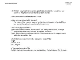
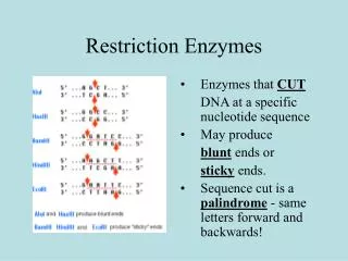
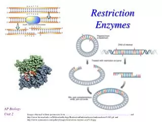
Introduction: • Restriction enzymes (also known as restriction endonucleases) found in bacteria (and harvested from them for use) and cut DNA up at specific sequences in the genome (blunt or sticky ends).
For example, the commonly used restriction endonucleaseEcoRIrecognizes every point in DNA with the sequence GAATTC, and cuts at the point between the Guanine and Adenine.
Usually, restriction enzymes only cut the DNA at or near a very specific nucleotide sequence known as arecognition site. • This “restrictive” nature of these enzymes allows molecular biologists to point exactly where the DNA is to be cut.
In many cases, the recognition site of a restriction enzyme consists of a palindromicsequence where the order of the bases is read the same on each side of the helix. • The two sides of the helix are antiparallel and the recognition sequence reads from 5’ to 3’ on either side of the helix.
Blunt ends are fragment ends of a DNA molecule that are fully base paired (no overhang), resulting from cleavage by a restriction enzyme. • Sticky ends fragment end of a DNA molecule with short single-stranded overhangs, resulting from cleavage by a restriction enzyme.
Whereas SmaI restriction enzyme cleavage produces "blunt" ends:
Nomenclature • Since their discovery in the 1970s, more than 100 different restriction enzymes have been identified in different bacteria. • Each enzyme is named after the bacterium from which it was isolated using a naming system based on bacterial genus, species and strain. • For example, the name of the EcoRI restriction enzyme was derived as shown in the box.
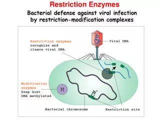
Restriction enzymes are part of a bacteria's ''immune'' system. • These are enzymes that cut DNA at specific sites (typically a four or a 6 base-pair sequence). • Bacterial DNA is modified to be protected by methylation while foreign DNA, such as incoming viruses, are not.
Usually, organisms that make restriction enzymes also make a companion modification enzyme ( DNA methyltransferase) that protects their own DNA from cleavage. • These enzymes recognize the same DNA sequence as the restriction enzyme they accompany, but instead of cleaving the sequence, they disguise it by methylating one of the bases in each DNA strand.
Thus, we can take a long piece of DNA and cut it with a restriction enzyme, generating numerous fragments. • Even a single-base change will destroy a restriction enzyme target site. • Likewise, even if a site is the same in two molecules, the length of DAN sequence between them may change. • Thus if two DNA molecules differ in sequence, they likely have different lengths for the fragments produced following treatment with restriction enzymes.
Endonucleases are enzymes that cleave the phosphodiester bond within a polynucleotide chain, in contrast to exonucleases, which cleave phosphodiester bonds at the end of a polynucleotide chain. • Typically, a restriction site will be a palindromic sequence four to six nucleotides long. Most restriction endonucleases cleave the DNA strand unevenly, leaving complementary single-stranded ends. These ends can reconnect through hybridization and are termed "sticky ends."
DNAs with the ECOR1 specific DNA sequence at different places will be cut into fragments of different lengths.
Procedure : • Warm your TE/DNA mixture at 55C for 15 -30 minutes. Pipet or light vortex to resuspend. • Prepare an enzyme digest of DNA by adding the required components to a clean microtube in the following order: • 14 µl H2O • 2 µl appropriate enzyme buffer (10X) • 3 µl DNA • 1 µl of enzyme (use BamHI, ClaI, EcoR, HaeIII, or HindIII)
3. Flick the tube to mix well and spin for 5 sec in the microfuge to bring all the components to the bottom. 4. Incubate at 37ºC in a water bath for two hours or overnight. 5. Run a gel (1% agarose gel as describe in Lab. 3 ) or freeze the samples until you have time to run the gel. 6. Record your observation.
Different restriction enzymes that recognize the same sequence are known as neoschizomers. These often cleave in different locales of the sequence. • Different enzymes that recognize and cleave in the same location are known as isoschizomers.