Download
1 / 34
350 likes | 512 Vues
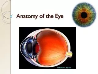
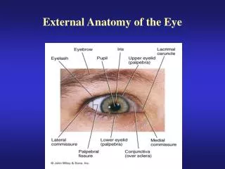
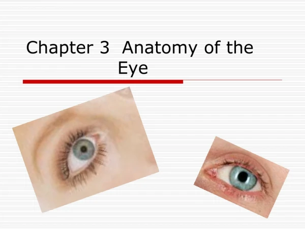
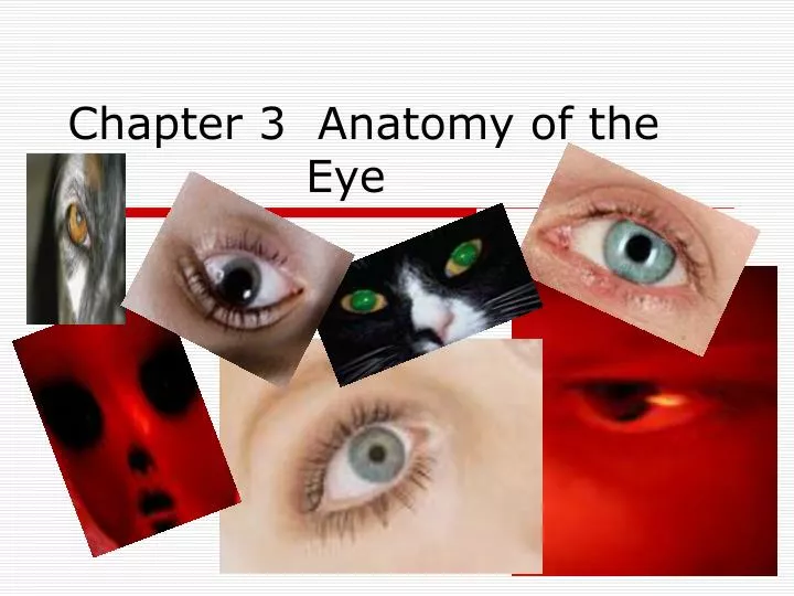
Chapter 3 Anatomy of the Eye. Sclera. The white part of the eyeball is called the sclera (say: sklair -uh). The sclera is made of a tough material and has the important job of covering most of the eyeball. Think of the sclera as your eyeball's outer coat.

E N D
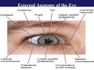
Sclera • The white part of the eyeball is called the sclera (say: sklair-uh). The sclera is made of a tough material and has the important job of covering most of the eyeball. Think of the sclera as your eyeball's outer coat.
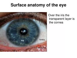
Look very closely at the white of the eye, and you'll see lines that look like tiny pink threads. These are blood vessels, the tiny tubes that deliver blood, to the sclera.
CORNEA • The part of the sclera in front of the colored part of the eye is called the cornea (say: kor-nee-uh). Unlike the rest of the sclera, which is white, the cornea is transparent, or completely clear, which lets light travel through it. The cornea helps the eye focus as light makes its way through. It is a very important part of the eye, but you can hardly see it because it's made of clear tissue. Like clear glass, the cornea gives your eye a clear window to view the world through
IRIS • Behind the cornea are the iris and the pupil. The iris (say: eye-riss) is the colorful part of the eye. When we say a person has blue eyes, we really mean the person has blue irises!
PUPIL • The iris is a muscle. This allows the iris to control how much light goes through the pupil (say: pyoo-pul). The pupil is the black circle in the center of the iris, and it lets light enter the eye. The pupils will get smaller when a light shines near them and they'll open wider when the light is gone.
Between the iris and cornea is the anterior (say: an-teer-ee-ur) chamber. This chamber is filled with a special transparent fluid that gives the eye oxygen, protein, and glucose (a type of sugar in the body) to keep it healthy.
TEARS • Our tears form a protective layer at the front of the eye and also help to direct the light coming into our eye.
After light enters the pupil, it hits the lens. The lens sits behind the iris and is clear and colorless. The lens' job is to focus light rays on the back of the eyeball - a part called the retina (say: reh-tin-uh).
Retina • Your retina is in the very back of the eye, past the vitreous body. Though it's smaller than a dime, it holds millions of cells that are sensitive to light. The retina takes the light the eye receives and changes it into nerve signals so the brain can understand what the eye is seeing.
The lens is suspended in the eye by a bunch of fibers. These fibers are attached to a muscle called the ciliary (say: sih-lee-air-ee) muscle. The ciliary muscle has the amazing job of changing the shape of the lens. That's right - the lens actually changes shape right inside your eye!
VITREOUS BODY • The biggest part of the eye sits behind the lens and is called the vitreous (say: vih-tree-us) body. The vitreous body forms two thirds of the eye's volume and gives the eye its shape. It's filled with a clear, jelly-like material called the vitreous humor. Ever touch toy eyeballs in a store? Sometimes they're kind of squishy - that's because they're made to feel like they're filled with vitreous humor. In a real eye, after light passes through the lens, it shines straight through the vitreous humor to the back of the eye.
Rods and Cones • The retina uses special cells called rods and cones to process light. Just how many rods and cones does your retina have? How about 120 million rods and 7 million cones - in each eye! • Rods and cones are most sensitive to yellow-green light.
RODS • Rods see in black, white, and shades of gray and tell us the form or shape that something has. Rods can't tell the difference between colors, but they are super-sensitive, allowing us to see when it's very dark.
CONES • Cones sense color and they need more light than rods to work well. Cones are most helpful in normal or bright light.
The retina has three types of cones - red, green, and blue - to help you see different ranges of color. Together, these cones can sense combinations of light waves that enable our eyes to see millions of colors.
I can’t see… • Sometimes someone's eyeball changes shape and the cornea, lens, and retina no longer work perfectly as a team. The person's eye may focus on what it sees in front of or behind the retina, instead of on the retina. When this happens, some of what the person sees will be out of focus.
Eye Glasses • To correct this fuzzy vision, many people, including many kids, wear glasses. Glasses help the eyes focus images correctly on the retina and allow someone to see clearly. As adults get older, their eyes change shape and they often need glasses to see things up close or far away. Most older people you know - like your grandparents - probably wear glasses.
To the Brain • Think of the optic nerve as the great messenger in the back of your eye. The rods and cones of the retina change the colors and shapes you see into millions of nerve messages. Then, the optic nerve carries those messages from the eye to the brain! The optic nerve serves as a high-speed telephone line connecting the eye to the brain.
Reflected light • Reaches the retina where it falls onto the cones and rods. • The critical part of the imaging process is the lens. • The lens gives the detailed information about the size, shape, and color of an object.
The lens is transparent with spherical surfaces. • It is convex which means thicker in the center.
FOVEA • The area near the center of the retina is called the fovea. • The detectors are packed tightly and details of the image are distinguished easily.
Peripheral vision • Light that enters your eye from the side does not fall on the fovea, but on the part of the retinal where there are fewer detectors. • This explains why peripheral vision is limited.
The placement and number of cones in your retina limit how well you see colors in your peripheral vision.
The placement of rods and cones differ in people which accounts for the diversity of vision.
Myopia • An eye that is too long or a cornea that is too steep causes myopia (or nearsightedness). In nearsighted eyes, the image isn't focused precisely inside the eye, causing blurring in the distance. The more nearsighted you are, the more blurred the distant object appears, and the thicker your glasses need to be. Most nearsighted people feel that their condition is severe, due to their dependence on glasses and contact lenses. In fact, only one in ten nearsighted individuals are actually in the "severe" or "extreme" categories.
Far-Sighted • Hyperopia • An eye that is too short, or a cornea that is not steep enough causes hyperopia (or Farsightedness). People with hyperopia see blurry when looking at close objects. Young people can slightly overcome hyperopia by using their focusing muscles to make the image clear. This gets harder as they get older. Currently, there are restricted options to correct hyperopia. Most operations are still under development.
The lens • The human eye can change the shape of the lens automatically. • The range of change that the lens can accomplish varies from person to person.