Download

1 / 44
460 likes | 1.3k Vues
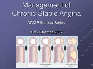
Chronic stable angina Guidelines . ACC/AHA 2002 Guideline for the Management of Patients With Chronic Stable Angina 2007 Chronic Angina Focused Update of the ACC/AHA 2002 Guidelines. Definition . Clinical syndrome characterized by discomfort in the chest, jaw, shoulder, back or arm

E N D
ACC/AHA 2002 Guideline for the Management of Patients With Chronic Stable Angina • 2007 Chronic Angina Focused Update of the ACC/AHA 2002 Guidelines
Definition • Clinical syndrome characterized by discomfort in the chest, jaw, shoulder, back or arm • Typically aggravated by exertion or emotional stress and relieved by nitroglycerin.
Canadian Cardiovascular Society Classification • Class I Ordinary physical activity does not cause angina, such as walking, climbing stairs. • Class II Slight limitation of ordinary activity. Angina on walking or climbing stairs rapidly, walking uphill, walking or stair climbing after meals • Class III Marked limitations of ordinary physical activity. Angina on walking one to two blocks on the level and climbing one flight of stairs • Class IV Inability to carry on any physical activity without discomfort—anginal symptoms at rest.
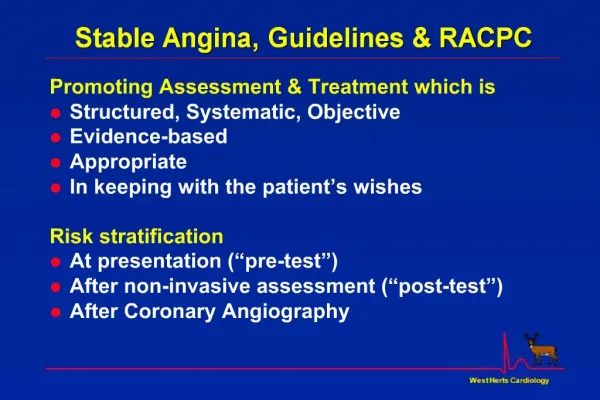
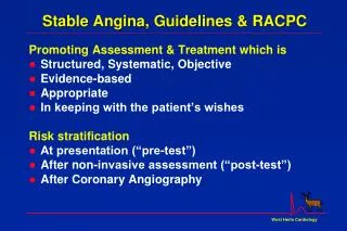
Purpose of diagnosis and assessment • Confirmation of presence of ischemia in patients with suspected angina • Identification associated conditions or precipitating factors • Risk stratification • To plan treatment options • Evaluation of efficacy of treatment
Exercise Electrocardiography • Most valuable for diagnosis when the patient's other clinical data suggest an intermediate probability of coronary disease • Uncertain value for patients with high or low pretest probability of coronary disease
CARDIAC STRESS IMAGING • Exercise stress testing is preferable to pharmacologic stress testing when the patient can exercise • Most useful for diagnosis in patients with an intermediate probability of disease. • Dobutamine perfusion imaging has significant limitations because it does not provoke as great an increase in coronary flow • Dobutamine is the agent of choice for pharmacologic stress echocardiography
Specific Patient Subsets • Treadmill electrocardiographic testing is less accurate for diagnosis in women than in men • Imaging technologies is also compromised by technical issues (e.g., breast tissue) in women • currently are insufficient data to justify replacing standard exercise testing with stress imaging in the initial evaluation of women.
EXERCISE TESTING for RISK ASSESSMENT AND PROGNOSIS in Intermediate or High Probability of CAD
EXERCISE TESTING for RISK ASSESSMENT AND PROGNOSIS in Intermediate or High Probability of CAD
Cardiac Stress Imaging as Initial Test for Risk Stratification of Patients with Chronic Stable Angina Who Are Able to Exercise
Cardiac Stress Imaging as Initial Test for Risk Stratification of Patients with Chronic Stable Angina Who Are Able to Exercise
Cardiac stress imaging as initial test for risk stratification of patients with chronic stable angina who are unable to exercise
ACC/AHA Guideline Criteria for Noninvasive Risk Stratification • High Risk (>3% Annual Mortality Rate) 1 Severe resting left ventricular dysfunction (LVEF < 0.35)2 High-risk treadmill score (score ≤ −11)3 Severe exercise left ventricular dysfunction (exercise LVEF < 0.35)4 Stress-induced large perfusion defect (particularly if anterior)5 Stress-induced multiple perfusion defects of moderate size6 Large, fixed perfusion defect with LV dilation or increased lung uptake7 Stress-induced moderate perfusion defect with LV dilation or increased lung uptake 8 RWMA (involving more than two segments) developing at low dose of dobutamine or at low heart rate (<120 beats/min)9 Stress echocardiographic evidence of extensive ischemia
ACC/AHA Guideline Criteria for Noninvasive Risk Stratification • Intermediate Risk (1%-3% annual mortality rate) 1 Mild or moderate LV dysfunction (LVEF = 0.35-0.49)2 Intermediate-risk treadmill score (−11 < score < 5) 3 Stress-induced moderate perfusion defect without LV dilation or increased lung intake4 Limited stress echocardiographic ischemia with a wall motion abnormality only at higher doses of dobutamine involving two segments or less • Low Risk (<1% Annual Mortality Rate) 1 Low-risk treadmill score (score ≥ 5) 2 Normal or small myocardial perfusion defect at rest or with stress* 3 Normal stress echocardiographic wall motion or no change of limited resting wall motion abnormalities during stress
Coronary Angiography for Risk Stratification in Chronic Stable Angina
Coronary Angiography for Risk Stratification in Patients with Chronic Stable Angina
Specific Goals for Risk Reduction Strategies in Chronic Stable Angina
Revascularization with Percutaneous Coronary Intervention and Coronary Artery Bypass Grafting in Stable Angina