Download
1 / 48
510 likes | 1.01k Vues
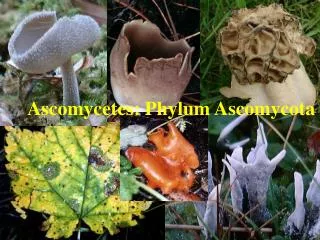
KULIAH 7. Ascomycota (continued) MIKROSKOPIK. YEAST, RAGI, KHAMIR… DLL. PEMBAGIAN ASCOMYCOTA. Class 1: Laboulbeniomycetes parasitic attachments to arthropods. Class 2: Protoascomycetes lack of ascogenous hyphae and ascomata . Class 3: Euascomycetes

E N D
KULIAH 7 Ascomycota (continued)MIKROSKOPIK YEAST, RAGI, KHAMIR… DLL
PEMBAGIAN ASCOMYCOTA Class 1: Laboulbeniomycetes parasitic attachments to arthropods. Class 2: Protoascomycetes lack of ascogenoushyphae and ascomata. Class 3: Euascomycetes most of the fungi which form ascomata. The orders are separated on the structure of the ascus and the manner of ascus opening.
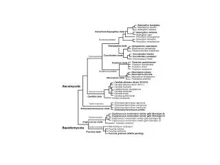
From Spatafora et al. 2006. A five-gene phylogeny of Pezizomycotina. Mycologia 98: 1018-1028
Filamentous ascomycetes Sacccharomycetales Characterized by DNA sequence analysis Archiascomycetes Basidiomycetes Ascocarps; ascogenous hyphae; specialized ascus tip; conidia; Woronin bodies Absence of ascogenous hyphae and ascocarps; most asci without specialized tips Classification from Alexopoulos et al. 1996
RAGI • Ascomycetous yeasts • Characterized by absence of ascogenoushyphae and ascocarps • Asci mostly prototunicate • Occur in slime fluxes, nectar, fresh or decaying fruit—able to grow in high osmotic conditions (high sugar content) • Others occur in soil, dung, water, digestive tracts of animals • Many species are symbiotic with insects
Somatic structures • Yeast • A single-celled fungus that reproduces by budding (or fission) • Pseudomycelium • Series of cells adhering after budding
Budding Bipolar Multilateral
Asexual reproduction • Conidia • Arthrospores
karyogamy plasmogamy budding copulation 2n somatic cells 1n somatic cells meiosis Mature ascus
Identification • Microscopic appearance • Unicellular or budding • Size & shape of yeast cells • Multilateral or bipolar budding • Form, structure and mode of ascus formation • Ascospore shape
Identification • Physiological tests—91 different tests • Ferment different sugars • Assimilation tests (carbon and nitrogen source) • Vitamin requirements • Growth at 37C • Growth in cyclohexamide • Hydrolyse urea • Form acid
Importance • Brewing • Baking • Food production • Industrial applications • Model systems (S. cerevisiae) • Probiotics • Animal pathogens
MICROSCOPIC (NON-YEAST)
Eurotium • Aspergillusanamorph • Cleistothecia yellow to orange-red • Wall composed of single layer of flattened cells • Ascosporesflattened, usually with equatorial groove. Ascospore by D. Geiser From Hanlin, 1998. Illustrated Genera of Ascomycetes Vol II
Emericella • Aspergillus anamorph • Cleistothecial wall surrounded by hülle cells • Ascospores small, colored, lens-shaped with flange From Hanlin, 1998. Illustrated Genera of Ascomycetes Vol II
Emericella Hülle cells, D. Geiser
Anamorphs--Aspergillus SEM by Charles Mims
Eupenicillium • Penicilliumanamorph • Cleisothecia hard, white becoming colored (yellow, orange, brown) • Ascospores small, hyaline or yellowish, lens-shaped From Hanlin, 1998. Illustrated Genera of Ascomycetes Vol II
Anamorphs--Penicillium phialides Branches (metulae)
Talaromyces • Paecilomyces or Penicilliumanamorph • Cleistothecium whitish to bright yellow • Wall composed of interwoven hyphae • Ascospores ellipsoidal, with spiny walls From Hanlin, 1998. Illustrated Genera of AscomycetesVol II
Anamorphs--Paecilomyces Divergent phialides with swollen base and long, tapering neck Colonies may be pink, purple, yellow, brown or white, but never green as in Penicillium spp.
The good and the bad • Penicillium spp.—antibiotic production • Penicilliumroqueforti—blue cheese • Penicillium spp.—blue and green molds on bread, cheese, fruits, vegetables • Aspergillus flavus—aflatoxins (moldy peanuts) • A. flavus/A. niger--aspergillosis
Penicillin • Penicillium notatum growing in Alexander Fleming’s Petri dish of Staphylococcus in 1928 led to the discovery of penicillin • Howard Florey & Ernest Chain (1939) began work on purification and trials • 1941—work moved to US (NRRL in Peoria, IL) to escape bombing in London (WWII) • Fermentation vessels and corn steep liquor • Mary Hunt (“Moldy Mary”) brought in P. chyrogenum on a melon • 1945—Fleming, Florey & Chain received Noble Prize
Penicillium notatum Penicillin prevents cross-linking of small peptide chains in peptidoglycan, the main wall polymer in bacteria. Newly formed cells are abnormal in shape and susceptible to osmotic lysis.
POWDERY MILDEW • Biotrophsof vascular plants • Biotroph: an obligate parasite growing on another living organism • 21 genera, 437 species infecting > 40,000 species of plants (mostly dicots) • Most species are host specific, a few are omnivorous, infecting hundreds of host species
Powdery Mildew Symptoms Photo by Claudia Nitschwitz
Characteristics • Mycelium is mostly superficial • Anchored to host epidermis by appressoria • Nutrients obtained via haustoria • Haustoria are intracellular structures • Overwinter as mycelium in infected buds or as ascomata • Asexual reproduction via conidia • Sexual reproduction via ascospores formed in cleistothecia
Asexual reproduction • Erect, hyaline conidiophores are usually formed on superficial mycelium; • One-celled, hyaline thin-walled conidia are produced holoblastically in basipetal chains • One colony can produce > 30,000 conidia
Conidia • Wind-dispersed • Germination can occur at low relative humidity • Germination involves germ tube, appressorium and penetration peg formation • Apex of penetration peg enlarges to form haustorium
appressorium Plasma membrane Penetration peg haustorium Host cytoplasm Plasma membrane Plant cell wall fungus
Sexual reproduction • Cleistothecia formed on superficial mycelium in late summer/early fall • Asci • Formed in basal layer • Globose to pyriform • Discharge of spore by rupture of ascus tip
Asci/Ascospores • One to numerous asci/cleistothecium • Ascospores hyaline, one-celled, ovoid • 1-8 ascospores/ascus • Number of asci/cleistothecium is important character in identification
Identification • Anamorph type • Number of asci/ascocarp • Cleistothecial appendages • Mycelioid • Rigid • Spear-like with inflated base • With curled tips • With dichotomously branched tips
Mycelioid Appendages • Several asci/ascocarp: • Eryisiphe(100 spp) • Oidiumanamorph • One ascus/ascocarp: • Sphaerotheca (50 spp.) • Appendages with curled tips • Oidiumanamorph
Dichotomously branched appendage tips • One ascus/ascocarp: • Podosphaera (12 spp.) • Oidiumanamorph • Several asci/ascocarp: • Sawadaea(6 spp.) • Oidiumanamorph
Spear-like appendages--Phyllactinia • Ovulariopsisand Streptopodiumanamorphs
Appendages with curled tips • Uncinula (81 spp) • Oidiumanamorph • Several asci/ascocarp