Download
1 / 41
410 likes | 744 Vues
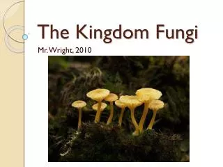
SRI CHUSNIATI. THE FUNDAMENTALS OF FUNGI. ORGANISM CLASIFICATION. Plantae: Seed plants, “paku-pakuan”, moss 2. Animalia: vertebrata & invertebrata 3. Protista *(procaryotic): ricketsia, bacteria, virus *(eucaryotic): algae, fungi, protozoa.

E N D
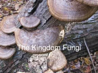
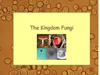
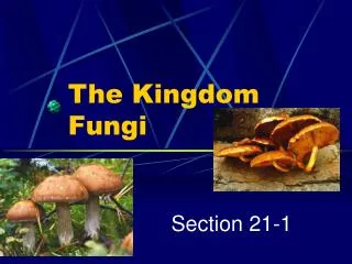
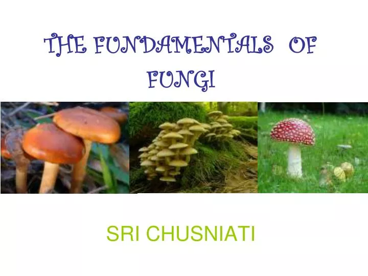
SRI CHUSNIATI THE FUNDAMENTALS OF FUNGI
ORGANISM CLASIFICATION • Plantae: Seed plants, “paku-pakuan”, moss 2. Animalia: vertebrata & invertebrata 3. Protista *(procaryotic): ricketsia, bacteria, virus *(eucaryotic): algae, fungi, protozoa
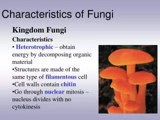
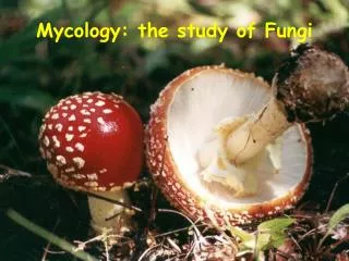
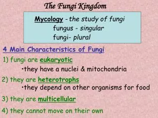
Algae : chlorophyl + • autotrophic • Fungi : chlorophyl - heterotrophic parasite, saprophyte Mycology Mycetes myces = fungi
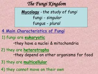
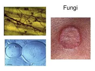
FUNGI / MUSHROOM *MACROFUNGI/ MACROMYCETES - mushroom *MICROFUNGI/ MICROMYCETES - yeast - mould Growth & developed on skin, hair, nail, mucous membrane, tissue animal & human # As an agent of caused to infection mycosis # It was produced of toxic metabolite Mycotoxicosis → Poison symptomatically
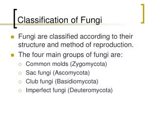
Purpose of classifications FUNGI A. Schizomycetes a. Actinomyces (anaerobe) Pseudomycetes b. Nocardia (aerobe) (false fungi) B. Mycomycetes (mucous fungus) C. Eumycetes (true fungi) 1. Phycomycetes (Zygomycetes & Oomycetes) hyphae non septate *2. Ascomycetes *3. Basidiomycetes *4. Deuteromycetes (fungi imperfecti) asexual spore, sexual spore ? Pathogenic generally * hyphae septate
MORPHOLOGY : • Difference with plant : • - chlorophyll – • - the composition of the cell wall was different • (chitin, glucan, cellulose, mannan) • - developed by spore • - trunk, branch, root, & leaves – • - function sharing of each part –
Fungi micro organism chemoheterotroph Nutrition Sources : • Carbon from organic materials • Nitrogen - organic (pepton) - an organic (ammonium & nitrate) • Mineral (P, K, Mg) macro element Fe, Zn, Cu, Mn, Mo micro element from substrate • Vitamin synthesis on their bioactivities / not from substrate ex/ thiamine & biotin • Water
Physical / Area Condition • Aeration : Kapang aerob Khamir aerob & faculttative anaerobe anaerobe lab CO2 inhibit of growth some fungi dimorphic morphogenesis and performing : macroconidia Trichophyton blastoconidia Histoplasma capsulatum
2. Light as a chemoheterotroph without light still growth - influence Spore perform (asexual & sexual)3. Temperature optimum 25-30o C psicrophilic, mesophilic, thermophilic
P 4. pH “Kapang”was optimum developed on acid pH pH 6-6,8 ; approximatelly at ranging : pH 2-8,5 Khamir was developed at pH 4-4,5 5. water activity (aw) Kapang < khamir < bacteria
Multicelluler Hyphae septate / not Aerob Cotton form / fibre Rhizoid / not place of rhizoid The Fungi was distinction on 2 groups : KHAMIR KAPANG • Mono/ unicelluler • Pseudohyphae / not • Aerob / anaerobe • The colony was pasta performed
KAPANGMORPHOLOGY Micellium + easy to be seen the growing white (at first) color according to the types of Kapang
PHYSIOLOGY • Water necessity (aw) to grow: kapang < khamir < bacteria • Temperature: mesophilic optimum 25o C - 30o C • Oxygen necessity & pH aerobic, pH 2,0 – 8,5 good acid pH • Nutrition: simple to complex amylase, pectinase, proteinase, & lipase enzyme production • Component inhibit: antibiotic Its to be slowest to growth, but already fast to growth if the inhibit component not active
The lucky “KAPANG” • Aspergillus oryzae: tape, soy sauce, tauco • Aspergillus niger: cytric acid, gluconat acid, amylase enzyme • Aspergillus wentii: pectinase enzyme • Auricularia polytricha: kuping mushroom • Mucor rouxii: saccarification process pati • Neurospora sitophila: red oncom • Penicillium notatum: penicillin • P. camemberti, P. roqueforti: cheese fragrant • Rhizopus oryzae, R. oligosporus: tempe, black oncom • Volvariella volvacea: merang mushroom
The unfortunately of KAPANG • Actinomyces israelii: Actinomycosis (teeth & tonsil) • Aspergillus niger: Otomycosis (tr. ear) • A. flavus: Aflatoxin • A. fumigatus: Aspergillosis (human & animal lung) • Blastomyces dermatitidis, B. brasiliensis: Blastomycosis • Candida albicans: Candidiasis (tr. Respiratorius, • tr. digestivus, tractus genitalia) • Coccidioides immitis: Coccidioidomycosis • Cryptococcus neoformans: Cryptococcosis • Histoplasma capsulatum: Histoplasmosis • Mucor mucedo: Food damaging • Nocardia astroides: Nocardiosis (human lung) • Trichopyton mentagrophytus: Tinea pedis (foot jaro)
MICOTOXIN *Aflatoxin (Aspergillus flavus): peas, corn, cereal *Eslanditoxin (Penicillium islandicum): rice *Patulin (Aspergillus clavatus): apple & apple products *Sterigmatosistin (Aspergillus versicolor, Aspergillus flavus) milk, grain, coffee, cheese *Tricotesen (Fusarium tricinchum): corn, cereal Toxin Symptom illness sometimes - fatal - carcinogenic - hallucinogenic
KHAMIRMORPHOLOGY • Macroscopic: like-bacteria colony • Size at ranging : length 1-5m to 20-50 m wide 1-10 m • Types of perform: circle, oval, cylinder, triangular, bottle, lemon, pseudohiphae, etc. • Cell formation: pseudohiphae/not CYTOLOGY Microstructure consists of capsule, cell wall bane, cytoplasm membrane, nucleus, vacuole, mitochondria, globule lipid, volutin/poliphosphat & cytoplasm
1. CAPSULE • Had by several khamir • Extra cellular component, mucous, cover up the outer part of cell wall bane • Polysaccharide and hetero polysaccharide • Hydrophobic 2. CELL WALL BANE Thin layer at immature cell at mature cell to be thicker generation time 1-6 hours - glucan/Cellulose 3-35 % - mannan 0-30 % - protein 6-8 % - variated chitin 0-2 % - lipid < 8-13,5 %
3. CITOPLASMA MEMBRANE • + 8 m thick • Consists of protein, ribonucleic acid & lipid • Nutrition transport & dismissal of metabolism product to outside 4. NUCLEUS • Surround by nucleus membrane (porous) • At fission/budding chromosome divided to 2 5. VACUOLA • Pocket contains translucent & aqueous fluid • > 1, size various
6. MITOCHONDRIA • P 0,4-0,6 m diameter 0,2-0,3 m • Respiration process 7. GLOBULA LIPID • Amount and various size 8.SITOPLASMA • Contains glycogen • Ribonucleic acid & protein (esp. in ribosome)
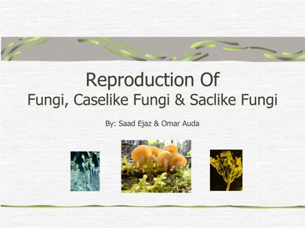
REPRODUCTION SYSTEM Some ways :1. budding 2. fission 3. bud fission vegetative reproduction 4. sporulation - asexual spore - sexual spore called generative reproduction • Cell budding duct formed from vacuole near nucleus cell wall bane. cell wall bane thinning protoplasm protruding goes out, and bigger (component is nucleus + cytoplasm) growth forming with new cells if size is almost = with the host goes separated or stay sticking & forming new bud
Categories of budding: • Multilateral: bud appear surround the tip of the cell at cylinder & oval formed cell • At all cell surface circle formed cell • Polar: just at one tip & Bipolar: at two tips lemon formed cell • Trigonopsis: bud at three tips of the cell (triangular form) • Pseudomicellium: if the bud don't liberated from its mother and continue budding
2. Cell fission • firstly, ‘bengkak/memanjang’ nucleus separated into 2 septa formed (2 layer) separated or formed into chain like mycelium • 3. bud fission • firstly, bud formed (where it sticks >) septa is formed separated • 4. Production of asexual spore: • arthrospore, blastospore, and klamidospore • 5. Production of sexual spore: • basidiospore and ascospore
PHYSIOLOGY CHARACTERISTICS • Grow well in enough water condition • Grow in medium with intense sugar or salt • aw 0,88-0,94 (osmophylic 0,62-0,65) Aw rice & cereal < 14 % • Optimal temperature 25o C – 30o C, max. temperature 35o C – 47o C some can grow at 0o C • pH 4,0 – 4,5 CLASSIFICATION & IDENTIFICATION • Morphology characteristics 1. vegetative reproduction 2. vegetative cell form, size, colour b. Culture characteristics: growing characteristics in liquid media, dense media c. Physiology characteristics d. Sexual reproduction
Khamir differ to 3 main classes: • Ascomycetes class: spore grow inside ascus • Basidiomycetes class: spore formed inside bacidium • Deuteromycetes class: not produce sexual spore called Fungi Imperfecti THE USAGE OF KHAMIR IN INDUSTRY Sacharomyces, Hansenula, Candida on making tape, brem Sach. cerevisae on making bread, beer, and wine
Dimorphic Fungi: fungi which has 2 phase that seen if grown at different temperature a. khamir phase at 37o C b. kapang phase at 24-28o C ex/ Sporothrix schenckii Histoplasma capsulatum Blastomyces dermatitidis Coccidioides immitis
Ascospore Basidiospore Zygospore Oospore SPORE ASEXUAL SEXUAL REPRODUCTION SYSTEMAsexual/vegetative : fission, budding, production of spore Sexual/generative : fusion of 2 nucleic • Sporangiospore • conidiospore • Arthrospore/oidospore • Klamidospore
1. Sporangiospore • Spore formed because cell protoplasm divide itself, formed small groups in sporangium pocket that placed on the tip of sporangiophore on hyphae has non septate. • Ex/ Rhizopus sp. Mucor sp.
2. CONIDIOSPORE -Spore formed because the tips of hyphae split. -Conidia formed at the tip of hyphae. -Pillar hyphae called Conidiophore. Ex/ Penicillium sp. Aspergillus sp.
3. ARTHROSPORE • Spore formed because a part of hyphae is broken & the wall thicken but not expand. ex/ Geotrichum Coccidioides Trichosporon
4. CLAMIDOSPORE • Spore formed because part of hyphae expand & create thick wall. • Rest phase • Many found at old hyphae. ex/ Candida albicans Epidermophyton
5. BLASTOSPORE • Spore which created from budding on yeast cell & the bud not liberated from its mother ex/ Rhodotorula sp. Blastomyces dermatitidis
1. ASCOSPORE • One-cell spore formed inside a pocket called ascus ex/ Saccharomyces
2. BASIDIOSPORE • Spore producted by basidia. Basidium exist on the tip of hyphae expanding that formed like vase/club ex/ Cryptococcus neoformans
3. ZYGOSPORE • Big thick-walled spore that formed if the tip of two swollen hyphae (gametangia) fuse (merged) ex/ Rhizopus Mucor
4. OOSPORE • Spore that formed inside oogonium because female gamet (oospher) fertilized by male gamet (antheredium) oospore • Inside each oogonium exist > 1 oospher
SUCCESSFUL HOPEFULLY GOODLUCK TO LEARN