Download
1 / 43
8.52k likes | 22.48k Vues
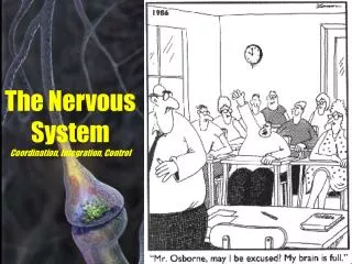
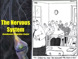
Control and Coordination. Some important terms. Stimulus- any external or internal factor that causes a living organism to react is called a stimulus. ( Pl: stimuli ) Response- the specific reaction shown by a living organism towards a stimulus

E N D
Some important terms • Stimulus- any external or internal factor that causes a living organism to react is called a stimulus. (Pl: stimuli) • Response- the specific reaction shown by a living organism towards a stimulus • Impulse- a electrical wave of excitation or irritation that travels across a neuron and carries specific messages.
Some important terms • Receptors- group of organs that receive sensory impulses and convey them to the brain • Effectors- group of organs that show responses to a specific stimulus
Neuron- • The fundamental structural and functional unit of the nervous system that carries impulses across the body.
Structure of a neuron A neuron is made up of two main parts; • Cyton • Axon Cyton – also called cell body, this part of neuron contains a large prominent nucleus in the center. The cell membrane is branched into several cytoplasmic branches called dendrites. Dendrites receive the impulses.
Structure of a neuron The cytoplasm is called neuroplasm. Inside the neuroplasm are scattered several stainable granules called Nissl granules. B) Axon – The long cytoplasmic projection of the neuron that extends from the cell body. An axon is covered by an insulating membrane called Neurolemna. Axons carry the impulse they receive from the cyton. They usually terminate into another neuron or an organ or gland or muscle.
Structure of a neuron In some neuron there is an additional envelop inside the neurolemna called the Myelin sheath which help in faster conduction of the impulse. Such neurons are called myelinated neurons and are mostly present in cerebral medulla. Impulse always travels from Cyton to Axon.
Types of Neurons • Sensory neurons- Those neurons that carry impulses from the receptors (sense organs)to the brain. These impulses are sensory in nature. • Motor neurons- Those neurons that carry impulses from the brain to the effectors . These impulses are motor in nature. • Associated neurons- They are also called mixed neurons as they carry impulses both to and from the brain.
Types of Neurons Motor Sensory Interneuron
Nerves A bundle of neurons with a common envelop around the axons called nerve membrane. A nerve provides a common pathway for the electrochemicalnerve impulses that are transmitted along each of the axons
Types of Nerves • Sensory nerves- Those nerves that carry impulses from the receptors (sense organs)to the brain. These impulses are sensory in nature. • Motor nerves- Those nerves that carry impulses from the brain to the effectors . These impulses are motor in nature. • Mixed nerves- They are also called mixed nerves as they carry impulses both to and from the brain. They are also called spinal nerves.
Classification of the Human Nervous System The human Nervous System is classified into three major divisions- The Central Nervous System The Peripheral Nervous System The Autonomous Nervous System
Central Nervous System • The CNS constitutes the main division of the Nervous system in Human Beings. • It comprises of two main organs concerned with control and coordination- • The Brain • The Spinal Cord.
The Brain • Location: Head region • Protection: a) Body protection made up of immobile bones that make up the skull. The skull is also called the Cranium. b) Membranous protection in the form of three membranes called the meninges. These three layers are called – Dura mater, Arachnoid mater and Pia mater. The cerebro-spinal fluid in between the three layers, protects the brain further from
Parts of the Brain Fore- Brain The fore brain makes up the largest part of the brain. The forebrain consists of the cerebrum, thalamus, and hypothalamus (part of the limbic system)
Fore Brain • Olfactory lobes: Two small lobe like structures situated in the lower part of the anterior brain. Controls the sense of smell
Fore Brain The cerebrum is the largest, most prominent part of the human brain. The longitudinal fissure partitions the cerebrum into right and left hemispheres, which are each separated into four lobes: • Frontal • Parietal • Temporal • Occipital • The cerebrum consists of the cerebral cortex (outer gray matter) and white matter. • The cerebral cortex is configured into convolutions (folds) that maximize surface area • The interior white matter consists of myelinated axons of neurons that link several regions of the brain
Lobes of the Cerebrum • Frontal Lobe- associated with reasoning, planning, parts of speech, movement, emotions, and problem solving • Parietal Lobe- associated with movement, orientation, recognition, perception of stimuli • Occipital Lobe- associated with visual processing • Temporal Lobe- associated with perception and recognition of auditory stimuli, memory, and speech
Fore Brain Thalamus is between the cerebral cortex and the midbrain, both in terms of its location and its neurological connections. Its function includes relaying sensation and special sense signals to the cerebral cortex, relaying motor signals from the cerebral cortex, and the regulation of consciousness, sleep and alertness.
Fore Brain Hypothalamus The hypothalamus is a portion of the brain that contains a number of small nuclei with a variety of functions. One of the most important functions of the hypothalamus is to link the nervous system to the endocrine system via the pituitary gland (hypophysis). The hypothalamus, is located below the thalamus, just above the brain stem The hypothalamus is responsible for certain metabolic processes and other activities of the Autonomic Nervous System. It synthesizes and secretes neurohormones
Mid Brain • It consist of Cruracerebri and corpora Quadregemina.
Hind Brain • Hind brain is made up of three parts: • Cerebellum • Pons • Medulla oblongata
Cerebellum • The hind part of brain. It is divided into cerebellar cortex and cerebellar medulla. • Cerebellar cortex has longitudinal fissures and not folds. While the medulla looks like a branched tree. • Cerebellum consists of 12% of the brain. Functions: • It controls muscular co-ordination • It regulates body balance.
Medulla Oblongata • The medulla contains the cardiac, respiratory, vomiting and vasomotor centers and deals with autonomic functions, such as breathing, heart rate and blood pressure
Pons Verolli • The ponsis a structure located on the brain stem. • The ponsrelayssensoryinformation between the cerebellum and cerebrum, aids in relaying other messages in the brain, controls arousal, and regulates respiration.
Spinal Cord- Structure • The spinal cord is an extension of the brain stem. • It runs mid-dorsally through the body and innervates the body. • The Sectional view of the spinal cord shows the outer white matter and inner grey matter. • The inter-neurons are present in the gray matter and help in reflexes. • Spinal cord is protected by a set of 33 bones called vertebral column and the three meninges.
Spinal Cord- Structure • A central canal is filled with the cerebro-spinal fluid. • 31 pairs of spinal nerves emerge out from either sides of the spinal cord. • Spinal nerves are mixed nerves as they have the sensory as well as the motor neurons in them.
Functions of the spinal cord • Spinal cord conducts impulses from the receptors to the brain as well as from the brain to the effectors. • Spinal cord controls reflexes.
Reflexes • A reflex is an involuntary, rapid response towards a stimulus with the active participation of the brain. Reflexes are protective responses against harmful stimuli. For ex: If we touch a hot plate we recoil our hand immediately to prevent it from getting burnt. This immediate and unconscious response is called reflex • Reflexes can be inborn or acquired.
Reflex Arc • The path travelled by an impulse during a reflex response is called a reflex arc. • A reflex arc begins from the receptors passes through the sensory neuron, passes via the interneuron to the motor neuron and then to the effectors.
PNS • The peripheral nervous system is composed of sensory neurons and the neurons that connect them to the nerve cord, spinal cord and brain, which make up the central nervous system. In response to stimuli, sensory neurons generate and propagate signals to the central nervous system which then processes and conducts signals back to the muscles and glands. • It is made up of 12 Pairs of Cranial Nerves and 31 pairs of spinal nerves.
ANS • The autonomic nervous system (ANS or visceral nervous system) is the part of the peripheral nervous system that acts as a control system functioning largely below the level of consciousness, and controls visceral functions. The ANS affects heart rate, digestion, respiration rate, salivation, perspiration, diameter of the pupils, micturition (urination), and sexual arousal. Whereas most of its actions are involuntary, some, such as breathing, work in tandem with the conscious mind.
Divisions of the ANS It is classically divided into two subsystems: the parasympathetic nervous system and sympathetic nervous system
Sympathetic Nervous System • Promotes a "fight or flight" response, corresponds with arousal and energy generation, and inhibits digestion. • Diverts blood flow away from the gastro-intestinal (GI) tract and skin via vasoconstriction. • Blood flow to skeletal muscles and the lungs is not only maintained, but enhanced (by as much as 1200% in the case of skeletal muscles).
Sympathetic Nervous System • Dilates bronchioles of the lung, which allows for greater alveolar oxygen exchange. • Increases heart rate and the contractility of cardiac cells (myocytes), thereby providing a mechanism for the enhanced blood flow to skeletal muscles. • Dilates pupils and relaxes the lens, allowing more light to enter the eye. • Inhibits peristalsis.
Parasympathetic Nervous System • Promotes a "rest and digest" response, promotes calming of the nerves return to regular function, and enhances digestion. • Dilates blood vessels leading to the GI tract, increasing blood flow. This is important following the consumption of food, due to the greater metabolic demands placed on the body by the gut. • The parasympathetic nervous system can also constrict the bronchiolar diameter when the need for oxygen has diminished.
Parasympathetic Nervous System • Normalizes Heart Beat levels. • During accommodation, the parasympathetic nervous system causes constriction of the pupil and lens. • The parasympathetic nervous system stimulates salivary gland secretion, and accelerates peristalsis, so, in keeping with the rest and digest functions, appropriate PNS activity mediates digestion of food and indirectly, the absorption of nutrients. • Is also involved in erection of genitals