Download
1 / 33
330 likes | 594 Vues
Apostolos I. Hatzitolios Associate Professor of Internal Medicine 1 st Propedeutic Department of Internal Medicine Department of Vascular Diseases and Hypertension Aristotle University of Thessaloniki, AHEPA Hospital Thessaloniki, Central Macedonia, HELLAS.

E N D
Apostolos I. Hatzitolios Associate Professor of Internal Medicine 1st Propedeutic Department of Internal Medicine Department of Vascular Diseases and Hypertension Aristotle University of Thessaloniki, AHEPA Hospital Thessaloniki, Central Macedonia, HELLAS Recent Guidelines for the Management of Arterial Hypertension
73% 70% 68% Diagnosis 59% 55% 54% 51% Treatment 34% 31% 29% 27% Regulation 10% NHANES II 1976-1980 NHANES III (Phase 2) 1991-1994 NHANES 1999-2000 NHANES III (Phase 1) 1988-1991 Diagnosis, regulation and treatment of hypertension in USA Hypertensive %
Guidelines 2007 • European Society of Hypertension • European Society of Cardiology Journal of Hypertension 2007;25:1105-1187
Stratification of CV risk in four categories Blood pressure (mmHg)
High/Very High Risk Subjects • BP ≥ 180 mmHg systolic and/or ≥ 110 mmHg diastolic • High systolic BP > 160 mmHgwith low diastolic BP (< 70 mmHg) • ≥ 3 cardiovascular risk factors • Diabetes mellitus or Metabolic syndrome • Hypertension Target Organ Damage orEstablished CV or renal disease
High/Very High Risk Subjects • One or more subclinical organ damages: • Electrocardiographic (particularly with strain) or echocardiographic (particularly concentric) LVH • Ultrasound evidence of carotid artery wall thickening or plaque • Increased arterial stiffness • Slight increase in serum creatinine • Reduced estimated glomerular filtration rate or creatinine clearance • Microalbuminuria or proteinuria • Established cardiovascular disease • Heart • Cerebrovascular • Renal • Peripheral artery • Ophthalmic disease
Appropriate BP measurement • Allow the patients to relax for several minutes in a quiet place • Take at leasttwo measurements spaced by 1-2 min and additional measurements if the first two are quite different [use phase I and V (disappearance) Korotkoff sounds to identify SBP and DBP] • Use a standard bladder but have a larger for fat arms and a smaller one for thin arms and children • Have the cuff at the heart level • Measure BP in both arms at first visit to detect possible differences due to peripheral vascular disease. In this instance, take the higher value as the reference one • Measure BP 1 and 5 min after assumption of the standing position in elderly subjects, diabetic patients and in other conditions in which postural hypotension may be frequent or suspected (e.g. heart, renal failure, SNS dysfunction, use of vasodilative agents) • Measure heart rate (at least 30 sec) after the second measurement in the sitting position
Home BP measurements • Self-measurement of BP at home is of clinical value, its prognostic significance is now demonstrated and these measurements should be encouraged in order to: • provide more information on the BP lowering effect of treatment at through and thus on the therapeutic coverage throughout the dose-to-dose time interval • improve patient’s adherence to treatment regimens • On the contrary, Self-measurement of BP should be discouraged when: • it causes anxiety to the patient • it induces self-modification of the treatment regimen
Ambulatory BP measurements • Although office BP should be used as reference, 24-h ambulatory BP monitoring may improve prediction of CV risk • Ambulatory BP should be considered, in particular, when: • considerable variability of office BP is found over the same or different visits • high office BP is measured in subjects otherwise at low CV risk • there is a marked discrepancy between BP values measured in the office and at home • there is a resistance to drug treatment • hypotensive episodes are suspected, particularly in elderly and diabetic patients • office BP is elevated in pregnant women and pre-eclampsia is suspected
BP thresholds (mmHg) for definition of Hypertension with different types of measurement
Particular conditions Isolated office hypertension (White coat hypertension) • Office BP persistently ≥ 140/90 mmHg • Normal daytime ambulatory or home BP < 130-135/85 Due to stress and SNS stimulation. CV risk is less than by raised office and ambulatory or home BP but may be slightly greater than by normotension Isolated ambulatory hypertension (Masked hypertension) • Office BP persistently normal (< 140/90 mmHg) • Elevated ambulatory (≥ 125-130/80 mmHg) or home BP (≥ 130-135/85 mmHg) CV risk is close to that of hypertension. Due to «normal» variationof circadian rhythm, autonomic nervous system dysfunction, physical or psychological stress, night consumption of alcohol, smoking and sleep apnea.
Guidelines for family and clinical history • Duration and previous level of high BP • Indications of secondary hypertension: • family history of renal disease (polycystic kidneys) • renal disease, urinary tract infection, haematuria, analgesic abuse (parenchymal renal disease) • drug/substance intake, such as: oral contraceptives, liquorice, carbenoxolone, nasal drops, amphetamines, steroids, non-steroidal anti-inflammatory drugs, erythropoietin, cyclosporine,cocaine (drug induced hypertension) • episodes of sweating, headache, anxiety, palpitation (phaeochromocytoma) • episodes of muscle weakness and tetany (aldosteronism)
Guidelines for family and clinical history 3. Risk factors: • family and personal history of hypertension and CV disease • family and personal history of dyslipidaemia • family and personal history of diabetes mellitus • smoking habits • dietary habits ; lack of physical exercise • obesity • snoring; sleep apnea (information also from partner) • Personality type; stress due to personal, family and environmental factors
Guidelines for family and clinical history 4. Symptoms of organ damage: • brain and eyes: headache, vertigo, transient ischemic attacks, sensory or motor deficit , impaired vision • heart: palpitation, chest pain, shortness of breath, swollen ankles • kidneys: thirst, polyuria, nocturia, haematuria • peripheral arteries: cold extremities, intermittent claudication 5. Previous antihypertensive therapy: • Drug(s)used, efficacy and adverse effects
Physical examination for secondary hypertension, organ damage and visceral obesity Signs suggesting secondary hypertension • Features of Cushing syndrome • Skin stigmata of neurofibromatosis (phaeochromocytoma) • Palpation of enlarged kidneys (polycystic kidneys) • Auscultation of abdominal murmurs (renovascular hypertension) • Auscultation of precordial or chest murmurs; Diminished and delayed femoral pulses femoral BP (aortic coarctation or aorticdisease)
Physical examination for secondary hypertension, organ damage and visceral obesity Signs of organ damage • Brain: murmurs over neck arteries, motor or sensory defects • Retina: fundoscopic adnormalities • Heart: location and characteristics of apical impulse, abnormal cardiac rhythms, ventricular gallop, pulmonary rates, peripheral oedema • Peripheral arteries: absence, reduction or asymmetry of pulses, cold extremities, ischemic skin lesions • Carotid arteries: systolic murmurs
Physical examination for secondary hypertension organ damage and visceral obesity Evidence of visceral obesity • Body weight • Increased body mass index [body weight (Kg)/height (m2)] overweight ≥ 25 Kg/m2; obesity ≥ 30 Kg/m2 • Increased waist circumference (standing position) ♂> 102 cm; ♀ > 88 cm
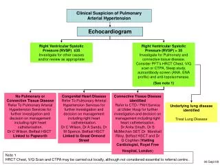
Laboratory investigations Routine tests: • Hemoglobin and hematocrit • Fasting plasma glucose • Fasting serum triglycerides • Serum total cholesterol, LDL-cholesterol, HDL-cholesterol • Serum creatinine, potassium, uric acid • Urinalysis (complemented by microalbuminuria dipstick test and microscopic examination) • Estimated creatinine clearance (Cockroft-Gault formula) or glomerular filtration rate (MDRD formula) • Electrocardiogram (ECG) • Thorax X-ray
Laboratory investigations Recommended tests • Echocardiogram • Carotid ultrasound • Quantitative proteinuria (if dipstick test positive) • Ankle-brachial BP index • Fundoscopy • Glucose tolerance test (if fasting plasma glucose > 5,6 mmol/l (102 mg/dL) • Home and 24h ambulatory BP monitoring • Pulse wave velocity measurement (where available)
Laboratory investigations Extended evaluation (domain of the specialist) • Further search for cerebral, cardiac, renal and vascular disease, mandatory in complicated hypertension • Search for suspected secondary hypertension suggested by history, physical examination or routine tests: • measurement of renin, aldosterone, • corticosteroids, • catecholamines in plasma and/or urine; • renal and adrenal ultrasound; • computer-assisted tomography (CT); • magnetic resonance imaging (MRI); • arteriographies
Searching for subclinical organ damage Importance of subclinical organ damage as an intermediate stage in the continuum of vascular disease and as a determinant of total CV risk. Heart • Electrocardiography should be part of all routine assessment of hypertensives in order to detect LVH, LV strain, ischemic condition and arrhythmias • Echocardiography is recommended whenever a more sensitive detection of LVH is considered useful. Concentric remodeling and hypertrophy carries the worst prognosis, while LV diastolic dysfunction, consists an early ECHO sign, which can be evaluated by Doppler measurement of transmittal velocities.
Searching for subclinical organ damage Blood vessels • Ultrasound scanning of extracranial carotid arteries is recommended in symptomatic carotid stenosis (previous TIA), but also in asymptomatic atherosclerosis suspected by carotid murmurs and reveals vascular hypertrophy, increased IMT, thickening of carotid bifurcation and presence of plaques. • Peripheral large artery stiffening (an important vascular alteration leading to isolated systolic hypertension in the elderly), can be measured by pulse wave velocity. This method might be more widely recommended if its availability were greater. • A low ankle-brachial BP index (<0,9) signals advanced peripheral artery disease
Searching for subclinical organ damage Kidney • Diagnosis of hypertension-related renal damage is based on a reduced renal function ordetection of hyperalbuminuria • Measurement of serum creatinine as well as estimation of glomerular filtration rate by specific formulas, should be part of routine procedures, allowing classification of renal dysfunction and respective stratification of CV risk • Presence of urinary protein should be sought in all hypertensives by dipstick. In dipstick negative patients, low grade albuminuria, namely microalbuminuria, should also be determined in spot urine and as ratio to creatinine excretion
Searching for subclinical organ damage Fundoscopy • Examination of eye grounds is recommended only in hypertensive with severe hypertension, since mild retinal changes (grade 1: arteriolar narrowing; grade 2: arteriovenous nipping) appear to be largely non-specific alterations except in young patients • In contrast, grade 3 (hemorrhages and exudates) and 4 (papilloedema) retinal changes, present only in severe hypertension and are associated with an increased CV risk
Searching for subclinical organ damage Brain • Silent brain infarcts, lacunar infarction (small / deep vessel disease), microbleeds and white matter lesions are not infrequent among hypertensives, especially elderly and can be detected by MRI or CT (MRI being generally superior to CT) • Availability and costs do not allow use of these techniques in asymptomatic patients • In elderly hypertensives, cognitive tests (e.g. Mini-mental scale) may also help to detect initial brain deterioration
Treatment of hypertension • Non pharmacological • Pharmacological
Goals of treatment • Primary goal of treatment is to achieve the maximum reduction in the long-term total risk of CV disease • This requires not only the treatment of raised BP per se, but also of all associatedreversible CV risk factors • BP should be reduced at least below 140/90 mmHg and even to lower values, if tolerated, in all hypertensive patients
Goals of treatment • Target BP should be at least < 130/80 mmHg in diabetics and in high or very high risk patients, such as those with associated clinical conditions, mainly stroke, myocardial infarction, renal dysfunction. Especially in proteinuria (<125/75 mmHg when >1gr/24h) • Despite the use of combination treatment, reducing SBP to <140 mmHg may be difficult and more so if the target is a reduction to <130 mmHg, moreover while a DBP reduction < 70 mmHg could be not beneficial. • Additional difficulties should be expected in elderly, obese and diabetic patients and in general, in patients with CV damage. In order to more easily achieve goal BP, antihypertensive treatment should be initiated before significant CV damage develops
Lifestyle changes • Lifestyle measures should be instituted whenever appropriate, in all patients, including those who require drug treatment, in order to lower BP, to control other risk factors and to reduce the number of doses of antihypertensive drugs to be subsequently administered • Lifestyle measures are also advisable in subjects with high normal BP (130-139 / 85-89)and additional risk factors to reduce the risk of developing hypertension • Lifestyle recommendations should not be given as lip service but instituted with adequate behavioral and expert support and reinforced periodically
Lifestyle changes Lifestyle measures widely recognized to lower BP or CV risk are: • smoking cessation • weight reduction (and stabilization) • physical exercise • reduction of salt intake • reduction of excessive alcohol intake • increase in fruit and vegetable intake and decrease in saturated and total fat intake (Mediterranean diet)
Lifestyle changes As long-term compliance with lifestyle measures is low and the BP response highly variable, patients under non pharmacological treatment should be followed-up closely to start drug therapy when needed and timely