Download
1 / 56
950 likes | 2.43k Vues
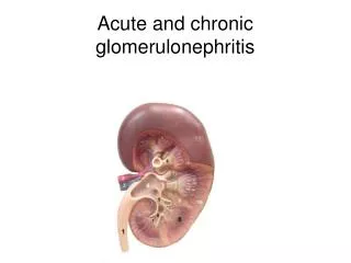
Glomerulonephritis. Dr Rodney Itaki Division of Pathology, SMHS, UPNG Anatomical Pathology Discipline. Gross anatomy. Ref: Goggle Images. Microanatomy. Robins Pathological Basis of Diseases, 6 th Ed. Figure 21.1. Glomeruli - Ultra filtration. Glomeruli & Renal Capsule. Blood Supply.

E N D
Glomerulonephritis Dr Rodney Itaki Division of Pathology, SMHS, UPNG Anatomical Pathology Discipline.
Gross anatomy Ref: Goggle Images
Microanatomy Robins Pathological Basis of Diseases, 6th Ed. Figure 21.1
Juxtaglomerular Apparatus +low BP & Ischaemia +Low NaCl
Pathophysiology of Glomerular Diseases • Types: • Immune or • Non-immune mediated injury
Immune mediated Glomerular Diseases • Immune mechanism can be of antibody-associated injury. Two forms are known: • Immune response resulting in injury due to deposition of soluble circulating antigen-antibody complexes in the glomeruli. Referred to asCirculating Immune complex injury. • Immune response resulting injury due to antibodies reacting in situ within the glomerulus. Referred to asCell Mediated Injury. • Others may be due to cytotoxic antibodies directed against the glomerular cells.
Non-immune Mediated Glomerular Diseases • 1. Metabolic glomerular injury. • Diabetic nephropathy: the glomerular lesion is glomerulosclerosis whereby there is thickening of the glomeular basement membrane. • 2. Hemodynamic glomerular injury. • This is due to the high intra-glomerular pressure caused by systemic hypertension or local change in glomerular hemodynamics (glomerular hypertension). • 3. Toxic glomerulopathies. • The toxic verotoxin from the E.Coli is directly toxic to renal endothelium and induces hemolytic-uremic syndrome in patients with infective diarrhea caused by E.Coli. Verotoxin interacts with specific cell membrane receptor inducing thrombotic microangiopathy.
Non-immune Mediated Glomerular Diseases • 4. Deposition disease. • There is deposition of abnormal proteins in the glomeruli inducing inflammatory reaction or glomerulosclerosis. For e.g. amyloidosis, cryoglobulins, light and heavy chain deposition disease. • 5. Infectious glomerulopathies. • Infectious microorganisms can cause injury by: • Direct infection of renal cell • Elaboration of nephrotoxins e.g. E.Coli • Intraglomerular deposition of immune complexes e.g. post-infectious glomerulonephritis. • Providing chronic stimulus for amyloidosis. • 6. Inherited glomerular diseases. • A common e.g. is: • Alport’s disease: Transmitted, as X-linked dominant trait. There is mutation in COL4A5 gene that encodes -5 chain of type IV collagen located on X-chromosome. The glomerular basement membrane (GBM) is affected.
The determinants of the severity of glomerular damage are • The nature of primary insult and secondary mediator system that evoke it. • The site of injury within the glomerulus. • The speed of onset, extend and intensity of disease.
Classification Glomerulonephritis Ref: Robins Pathological Basis of Diseases, 6th Ed. Table 21.3
Primary Glomerulonephritis Ref. Robins Pathological Basis of Diseases, 6th Ed. Figure 21.29
Clinical Presentation • Nephrotic Syndrome • Nephritic Syndrome • Others – mixture of symptoms of nephrotic & nephritic syndrome
Diagnostic Criteria - NephroticSyndrome • Increased BM permeability – increased urinary loss of plasma proteins esp. albumin • Massive Proteinuria - >4grams per day. 24 Hr Urine. • Hypoalbuminemia – from proteinuria. Serum concerntration of <3gm/100ml. • Generalised oedema – decreased plasma colloid oncotic pressure. • Hyperlipidemia & hypercholesterolemia – increased hepatic lipoprotein synthesis.
Diagnostic Criteria - Nephritic Syndrome • Inflammatory rapture of glomerular capillaries resulting in bleeding into urinary space (Bowman’s capsule). Proteinuria and oedema mild. • Oliguria – Reduced GFR causing reduced urine output. • Azotemia – elevated BUN. • Hypertension – increased fluid retention & Renin-Angiotension-Aldosterone activation by ischaemic kindneys • Hematuria – form RBC casts & granular casts
Disorders Manifest as Nephrotic Syndrome Minimal Change Disease (Lipoid nephrosis) Focal Segmental glomerulosclerosis Membranous glomerulonephritis Diabetic nephropathy Renal amyloidosis Lupus nephropathy
Minimal Change (lipoid nephrosis) Disease • Common cause of nephrotic syndrome in children. • Immune mediated • Characterized by loss of foot processes of epithelial cells (podocytes) in glomeruli. • Responds to steroid therapy
Minimal Change Disease (Lipoid Nephrosis) Visceral epithelial cells show uniform and diffuse effacement of foot process Thin BN. No proliferation
Minimal Change Disease Normal glomerular tuft. No hypercellularity. Thin BM. Ref: www.kidneypathology.com
Focal Segmental Glomerulosclerosis • Clinically similar to minimal change disease but occurs in older patients • Can occurs as primary or secondary disorder. • Primary – idiopathic focal segmental glomerulosclerosis • Secondary – HIV, heroine addiction, sickle cell disease, IgA nephropathy, certain forms of inherited nephrotic syndrome.
Idiopathic FSG • 10-15% of nephrotic syndrome in adults and children. • Higher incidence of hematuria, reduced GFR and hypertension • Non-selective proteinuria • Respond poorly to steroid therapy • Many progress to chronic GN & 50% develop end stage renal disease in 10 years • Immunoflurescence microscopy shows IgM & C3 in sclerotic glomerularsegments
Focal Segmental GlomerularSclerosis • Sclerotic segment shows deposition of hyaline masses • Lipid in sclerotic area (small vacuoles) Foam cells Ref:www.med.niigata-u.ac.jp
Membranous Glomerulonephritis • Most common cause of nephrotic syndrome in adults (40%). • Characterised by: diffuse thickening of glomerular capillary wall & accumulation of electron-dense immunoglobulin containing deposits along epithelial and subepithelial side of BM
Membranous Glomerulonephritis • Primary – 85% of cases no association with any condition. • Secondary – association with drugs (e.g. NSAIDS), tumors (e.g. CA lung and colon), SLE (15% of GN), infections (e.g. chronic Hep B, C, malaria, syphilis, schistosomiasis) & metablic disorders (e.g. DM & throiditis)
Membranous Glomerulonephritis • Primary – Auto-Immune disorder caused by antibodies to renal autoantigen. • Secondary – chronic antigen-antibody mediated disorder. • Activation of compliment pathway cause injury to capillary wall and cause increased protein leakage.
Membranous GN Diffuse thickening of capillary wall without increase in number of cells Diagrammatic representation Ref: Robins Pathological basis of Diseases, 6th Ed. Fig. 21.19
Diabetic Nephropathy Characterised by: • BM markedly thickened. • Diffuse of nodular mesangial accumulations of BM like material.
Diabetic Nephropathy Ref: Robins Pathological Basis of Diseases, 6th E. Table 20.1
Diabetic Nephropathy • Capillary BM thickening. • Diffuse glomerulosclerosis. • Nodular glomerulosclerosis. Ref: www.unckidneycentre.org
Basement membrane Thickening Thickened BM Ref: www.intechopen.com
Renal Amyloidosis • Deposition of amyloid protein in glomeruli. • Early stage present as Nehprotic Syndrome. • End stage – Chronic renal failure.
Amyloidosis Deposition of abnormal protein in the glomerulus & blood vessel wall Amyloid deposits
Amyloidosis Congo red stain. Examined under polarization microscopy. “Apple-green” birefringence. Ref: www.pathology.vcu.edu
Lupus Nephropathy • SLE – auto-immune disorder. • Lupus Nephropathy is renal component of SLE. • Immune Complex disposition in subendothelial location causing immune mediated injury to glomeruli. • May have features of Nephritic syndrome as well • Histologically: various forms. None specific for SLE
Disroders Manifest By Nephritic Syndrome Poststreptococcal GN Rapidly Progressive (crescentic) GN Goodpasture syndrome Alport Syndrome
Poststreptococcal GN (Acute Proliferative GN) • Immune mediated. • Sequelae of nephritogenic strains of Group A beta-hemolytic streptococcal skin infection or pharyngitis. • Deposition of immune complexes in glomeruli cause inflammation and damage BM. • Common in children age 6-10 yrs.
Post-streptococcal GN Normal glomerulus Acute proliferate GN Hypercellularity due to intercapillary leucocytes & proliferation of glomerularcells (mesangial cells, endothelial cells) Ref: Robins Pathological Basis of Diseases, 6th Ed. Fig 21.16
Rapidly Progressive (crescentic) GN • A syndrome and not a specific diagnosis. Immune mediated. • 3 types – Type I (ANCA Neg, Anti glomerular BM antibodies), II (Immune complex) & II (ANCA Pos pauci-immune form). • Can occur as primary or in association with other diseases. • Disease association: E.g. Goodpasture syndrome (Type I), SLE (Type II) Wegener granulomatosis (Type II).
Rapidly Progressive GN • Clinically can progress rapidly to renal failure in weeks or months. • 50% poststreptococcal with immune complex deposition. • 10% due to antiglomerular BM antibodies (Type I, ANCA Neg) and present as clinically as Goodpasture syndrome.
Rapidly Progressive (Crescentic) GN Ref: www.geekymedics.com
RPGN or Crescent GN Collapsed glomerular tufts Mass of crescent shaped proliferating cells & leucocytes Ref: Robins Pathological Basis of Diseases, 6th Ed. Fig 21.17
Goodpasture Syndrome • Also known as Antiglomerular BM disease • Immune mediated: Antibodies directed against antigens in glomerular and alveolar BM • Clinically present as Nephritic syndrome & pneumonitis with hemoptysis • Peak incidence in male in mid 20s • Histology shows RPGN crescentic morphology with linear immunofluorescence staining.
Alport Syndrome • Hereditary nehpritis with nerve deafness & ocular disorders (lens dislocation & cataracts) • Inherited disorder. Heterogenous mode of inheritance. Most patients have X-linked dominant pattern. • Genetic basis: mutation in gene for alpha 5 chain of type IV collagen resulting in defective GBM synthesis. • Symptoms appear between 5-20 yrs and renal failure by 20-50 years. • Histology: irregular BM thickening with foci of splitting of the lamina densa
Other Glomerular Disorders (Not Nephrotic or Nephritic IgA Nephropathy (Berger Disease) Membranoproliferative GN
IgA Nephropathy (Berger Disease) • Very common entity • Defined by deposition of IgA in the mesangium • Frequent cause of recurrent gross or microscopic hematuria. • Can occur as Primary or secondary to other disorders (liver and celiac disease) • Affects children and adults • Slow progression to chronic renal failure occurs