Download
1 / 35
350 likes | 353 Vues
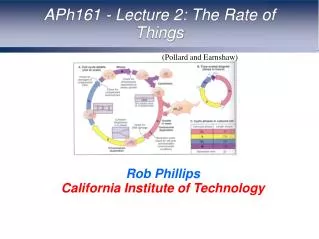
BE/APh161 – Physical Biology of the Cell. Rob Phillips Applied Physics and Bioengineering California Institute of Technology. Ion gating driven by ligands. Ligand-gated channels. Ion gated channels: Acetylcholine. Data for the gating of nicotinic acetylcholine receptor.

E N D
BE/APh161 – Physical Biology of the Cell Rob Phillips Applied Physics and Bioengineering California Institute of Technology
Ion gating driven by ligands • Ligand-gated channels.
States and weights for binding problems • We work out the probability of the binding probability by making a model of the solution as a lattice.
Binding curves and binding free energy • These simple binding curves illustrate the way in which the binding probability depends upon the Kd or the binding energy.
Exploring Promoter Architecture: Can We Compute How Cells Decide?
Exploring Promoter Architecture: Can We Compute How Cells Decide?
Where we are headed: Can We Compute How Cells Decide? Bintu et al. (2005)
Some other examples • Data and fits using our binding formula.
Gibbs’ second law • One idea only: to find the privileged terminal state of a system, maximize the entropy. • A corollary: minimize the free energy – this is for a system in contact with a heat bath. • My point here is to get us all to think about the chemical potential.
The gibbs distribution System in contact with an energy reservoir System in contact with a particle and energy reservoir Probability for finding the system in microstate i: Probability for finding the system in microstate i: Boltzmann distr. - partition f. Gibbs distr. grand partition f. res.controls av. # of particles <N> in the syst. Treservoircontrols av.energy <E> of the system
ligand-receptor binding: State variable description • Consider a single receptor in contact with the surrounding heat bath and particle reservoir. • Two-state (b/u), is an indicator of the state of binding • The energy is • Evaluate aver. # of ligands bound, <N>: favorable interaction btw L and R Contact with a particle reservoir Contact of the system with a thermal reservoir • Recall that the chem.potential of an ideal solution is => is the energy difference upon taking the ligand from solution and placing it on the receptor can also be computed as
Cooperativity and binding • Interestingly, many (if not most) of the real world binding problems we care about in biology do not satisfy the simple binding model (sometimes called the Langmuir adsorption isotherm) we have worked out so far. • The classic example (i.e. the hydrogen atom of binding problems) is hemoglobin.
Hemoglobin as a case study in cooperativity • Hemoglobin - the classic example of ligand-receptor binding • Cooperativity: the binding energy for a given ligand depends upon the # of ligands that are already bound to the receptor • Intuitively: conformational change upon binding => the next ligand experiences a different binding energy The protein hemoglobin: 4 polypeptide chains (2 -chains, 2 -chains), each carries a heme group => protein can bind up to 4 molecules of O2 several 100s hemoglobin molecules apps.uwhealth.org Oxygen binds to heme on the hemoglobin molecules The heme group includes a porphyrin ring (gray line) + iron
Hemoglobin as a case study in cooperativity • Hemoglobin-oxygen binding: language of two-states occupation variables. State of system is described with the vector where i: i = 0 (unbound), i = 1 (bound) • Q.: what is the average # of bound O2 molecules as a function of the O2 concentration(or partial pressure)? A toy model of a dimoglobin • To illustrate the idea of cooperativity: imagine a fictitious dimoglobin [=dimeric hemoglobin] molecule which has 2 O2 binding sites (e.g., clams) • => 4 distinct states • The energy of the system: measure of the cooperativity Energy associated with O2 being bound to one of the 2 sites
A toy model of a dimoglobin • The grand partition function (sum over the 4 states): • => compute the probabilities for each classes of states: unoccupied, single occupancy, double occupancy Single occupancy Both sites occupied Parameters used: =–5 kBT, J= –2.5 kBT, c0 = 760 mmHg
Talking across the membrane • Membrane proteins are characterized in some cases by transmembrane alpha helices and cytosolic domain that passes along the signal.
Coupling receptors to enzyme action • Receptor binding changes the probability of the “active” state.
Doing work to change the protein state • A wonderful and important topic for our consideration is that of posttranslational modifications. • One of the tricks performed by the cytoplasmic side of a receptor (or its partners) is to do some posttranslational modification.
phosphorylation • In bio systems, changes in envir.conditions => the activity of an enzyme must be rapidly altered • One of the most important regulatory modes in all of biology: regulation of protein activity by covalent attachment of phosphate groups • The substrate for protein phosphorylation: target protein and ATP • The enzyme: protein kinase (transfers the terminal phosphate group from ATP to a chemical group on a protein) • A phosphate group carried 2 “-” charges => causes a dramatic change in the local charge distribution on the surface of the protein => drastic, large scale effect on protein structure and ability to bind • This alteration is reversible: protein phosphatase
The diversity of kinases • “The whole molecular control network, leading from the receptors at the cell surface to the genes in the nucleus, can be viewed as a computing device; and, like that other computing device, the brain, it presents one of the hardest problems in biology.” • Catalytic domains shown in green Roughly 250 aa long.
phosphorylation: two internal state variables • What is the fraction of activated proteins? How does it depend on the state of phosphorylation? • Model: The “structural” state of the protein (active/inactive): s: s = 0 => inactive, s = 1 => active The state of phosphorylation of the protein: p: p = 0 => unphosphorylated, p = 1 => phosphorylated • The state of phosphorylation can alter the relative energies of the active and inactive states => at equilibrium, most of the phosphorylated molecules will be in active form • I1 and I2 are the electrostatic interaction energies btw the two charges in the active and inactive states
phosphorylation: two internal state variables • Using the variables, the free energy of the protein is which simplifies to => states&weights:
phosphorylation: two internal state variables • From the states and weights: Probability of the protein being in the active state, if it is notphosphorylated Probability of the protein being in the active state, if it is phosphorylated • The change in activity due to phosphorylation:
phosphorylation: two internal state variables • In the toy model in the figure, => -increase in activity upon phosphorylation • In the cell, the increase in activity upon phosphorylation spans from factors of 2 to 1000.
Eukaryotic signal transduction • A more precise realization of the implementation of signaling. • We begin with an example that is simple both conceptually and mathematically, namely, prokaryotic two-component signal transduction..
Two-Component Signal Transduction • Next few slides are courtesy of Michael Laub (MIT) and Mark Goulian (Upenn) – experts in the quantitative dissection of signaling networks. • This figure shows the generic features of the two-component signal transduction systems.
SprE FimZ HydD RcsC NtrB QseC DpiA RcsD KdpD PhoQ UhpB NarQ PhoR CpxA DcuS NarX CusS EnvZ RstB BasS ArcB TorS TorR RstA BasR PhoB KdpE NarL PhoP CusR UhpC DcuR NarP OmpR CpxR RcsB OmpR DpiB NtrC UvrY CheY YedW HydG YpdB QseB YehT CheB YfJR CreB CheA CreC BaeR YpdA YehU AtoC YfhK BaeS AtoS Coordinating multiple signaling systems in a single cell EvgA EvgS BarA YedV animation by Mark Gouilan
(use complete set of purified RRs) HK + ATP* HK~P* + ADP ......... - RR1 RR2 RR3 RR44 Phosphotransfer profiling incubate, separate by SDS-PAGE HK~P* + RR HK + RR~P* HK~P RR~P
C. crescentus PhoR profile – 5 min phosphotransfer reactions +PhoB Assessing Specificity: Phosphotransfer Profiling C. crescentus PhoR profile – 60 min phosphotransfer reactions +PhoB • histidine kinases exhibit a strong kinetic preference in vitro • for their in vivo cognate substrate • specificity based on molecular recognition
Signal integration • Once we finish with our concrete example of chemotaxis, we will turn to the way in which cells decide where to put new actin filament and that will make us face this question of signal integration.