Download
1 / 49
590 likes | 1.12k Vues
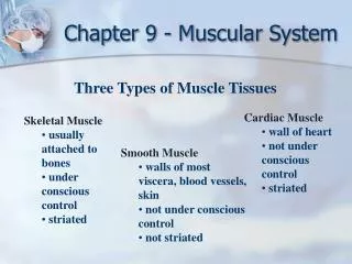
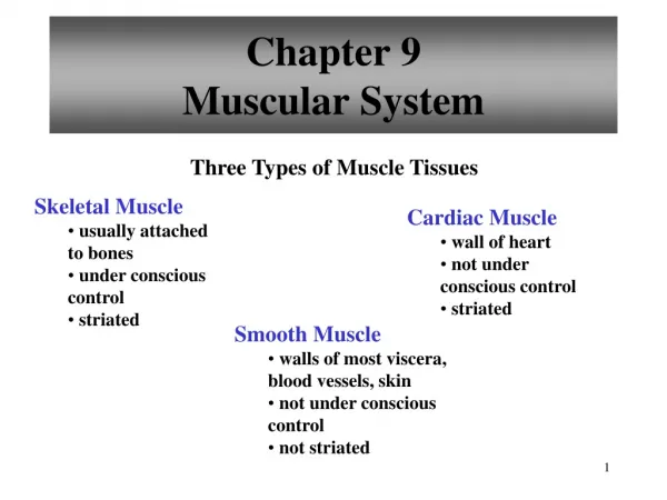
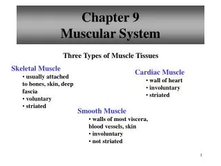
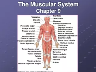
Chapter 9 The Muscular System. Skeletal Muscle Structure. Tendon – connect muscle to bone Fascia – outermost covering; covers entire muscle & continuous w/tendon; separates muscle from adjacent muscles Aponeuroses - connect muscle to muscle. Skeletal Muscle Structure.

E N D
Skeletal Muscle Structure • Tendon – connect muscle to bone • Fascia – outermost covering; covers entire muscle & continuous w/tendon; separates muscle from adjacent muscles • Aponeuroses- connect muscle to muscle
Skeletal Muscle Structure Coverings: • Epimysium – covers entire muscle (under fascia) • Perimysium – covers muscle bundle (fascicle) • Endomysium – covers each fiber (cell) • Sarcolemma – cell membrane
Skeletal Muscle Structure Skeletal Muscle Structure – Cont. • Sarcoplasmic reticulum (SR) channels for transport • Myofibrils – threads that compose muscle fibers; contain protein filaments: 1. actin – thin 2. myosin – thick
Muscle Fiber(muscle cell) • Cisternae of SR – enlarged portions • Transverse tubules (T-tubules) – important in muscle contraction • Sarcoplasm – cytoplasm
Parts of a Sarcomere • Z lines – end points • M line – middle • I band – on either side of Z line; actin filaments only • H zone – on either side of M line; myosin filaments only • A band – overlapping actin & myosin filaments
Neuromuscular Junction – junction b/t motor neuron & muscle • Motor neuron – carries impulse from brain or spinal cord to muscle • Motor end plate – end of muscle fiber; many nuclei & mitochon- dria located here
Neuromuscular Junction • Neurotransmitters (ntm) chemicals that help carry impulses • Motor unit – 1 motor neuron & fibers that it stimulates • Synaptic vesicles – store neurotransmitter; most common – acetylcholine (ACh)
Neuromuscular Junction Animation • Neuromuscular Junction Animation
Troponin & Tropomyosin • 4 proteins are found in muscle cells: actin, myosin, troponin & tropomyosin • troponin – appear as globules; provide a binding site for Ca+² • tropomyosin– appear as ribbons; cover the myosin cross-bridge binding sites in a relaxed muscle
Sliding Filament Theory (How Muscles Contract) • Muscle fiber stimulated by release of ACh from synaptic vesicles of neuron • ACh causes impulse to travel to muscle cell membrane • Transverse tubules (T-tubules) carry impulse deep into muscle fibers • Sarcoplasmic reticulum releases Ca ions (Ca²+) • Ca²+ bind to troponin, tropomyosin moves, exposing binding sites on actin filaments
Cross Bridge Animation • cross bridge animation
Sliding Filament Theory (How Muscles Contract ) • Linkages form b/t actin & myosin • Actin filaments move inward, shortening the sarcomere • Muscle fiber relaxes when Ca²+ are transported back to S.R. • The enzyme cholinesterase (or AChesterase) decomposes ACh
Sliding Filament Theory • Relaxed muscle – binding sites on actin are covered by tropomyosin
Sliding Filament Theory • Ca²+ binds to troponin • Tropomyosin slides out of the way • Myosin binds to actin & pulls inward • Sarcomeres shorten & muscle contracts
Sliding Filament Animation • sliding filament animation
Energy for Muscle Contraction • ATP (adenosine triphosphate) provides the energy for muscle contraction • When ATP is converted to ADP(adenosine diphosphate) by losing the last phosphate, energy is released.
Energy for Muscle Contraction • Cells depend on cellular respiration of glucose to synthesize ATP • An additional source is creatine phosphate
Energy for Muscle Contraction • Creatine phosphate stores excess energy • Can be used to convert ADP back into ATP • Anaerobic respiration (in the absence of O2) provides few ATP’s, while aerobicresp. (in the presence of O2) provides many ATP’s
Creatine Phosphate High amts. of ATP - ATP is used to Low amts. of ATP – CP is used synthesize CP, which stores energy to resynthesize ATP. for later use.
Importance of Myoglobin • l.a. carried by blood to liver; liver can convert l.a. to glucose, but requires ATP (ATP being used for muscle contraction) • myoglobin – stores O2 in muscle cells; gives muscle its red color
Aerobic vs. Anaerobic Respiration Carried by blood to liver; liver can convert l.a. to glucose, but requires ATP (ATP being used for muscle contraction) Imp. b/c blood supply during muscle contr. may decrease As l.a. accumulates, O2 debt occurs
Oxygen Debt • Strenuous exercise leads to O2 deficiency & lactic acid buildup • ATP provides energy for muscle contraction • Amt. of O2 needed to convert accumulated l.a. to glucose & restore ATP levels = O2 debt • L.A. accumulation leads to muscle fatigue b/c pH of muscle cell is lowered & muscle cannot contract
Muscle Cramp • Muscle cramp – fatigued muscle has lack of ATP needed to move Ca+² back into S.R.; cross bridges not broken • Rigor mortis – takes up to 72 hrs. to occur; sarcolemma becomes more permeable to Ca+² & ATP levels insufficient
Myogram Pattern or graph of a muscle contraction A single contraction is called a muscle twitch 3 parts: Latent (lag) phase – brief pd. of delay b/t when the stimulus is applied & actual contraction occurs Contraction Relaxation – return to original state
Patterns of Contraction • a) MuscleTwitch – single contraction • b) Staircase Effect many stimuli closely spaced w/complete relaxation in b/t; each contraction generate incr. force
Patterns of Contraction • c) Summation – when the 2nd stimulus occurs during the relaxation pd. of 1st contr.; the 2nd contr. generates more force • d) Tetany – when twitches fuse into 1 sustained contr.
Muscle Facts • If a muscle is stimulated twice in quick succession, it may not respond the 2nd time – called refractory period • Threshold – the minimum stimulus needed to cause a contraction • All-or-none – increasing the strength of the stimulation does NOT incr. the degree of contraction (a muscle contracts completely or not at all)
More Facts • Incr. stimulation from motor neurons causes a greater # of motor units to contract & vice versa • Called recruitment of motor units • Incr. the rate of stimulation also incr. the degree of contraction • Muscle tone – a sustained contraction caused by nerve impulses from s.c. to a small # of muscle fibers in the back, neck, etc.; maintains posture
Origin & Insertion • Origin – end of muscle that attaches to stationary bone • Insertion – end of muscle that attaches to moving bone • During contr., insertion is pulled toward origin
Muscle Functions in Groups Prime mover – responsible for most of the movement (ex.- biceps) Synergist – aids the prime mover Antagonist – resists the prime mover & causes movement in the opposite direction (ex. - triceps)
Levers • Parts of a lever: • wt., force, pivot • 3 types of levers: • 1st class – W-P-F • (seesaw/scissors) • 2nd class – P-W-F • (wheelbarrow) • 3rd class – W-F-P • (forceps)
Bones & Muscles as Levers • Forearm bends – 3rd class lever (biceps attaches at a pt. on the radius below the elbow joint) • Forearm straightens - 1st class lever ((triceps attaches at a pt. on the ulna • above the elbow joint)
Bones & Muscles as Levers Standing on tip-toe – 2nd class lever (P-W-F)