Download
1 / 40
420 likes | 433 Vues
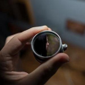
Eye Injuries and Illnesses. Anatomy of the Eye. Eye Injury. Chemical Burns. Treatment should be immediate, even before making vision tests! Premedicate with proparacaine or tetracaine. Copious irrigation: LR or NS X 30 min. Wait 5 minutes and check pH. If not normal, repeat.

E N D
Chemical Burns • Treatment should be immediate, even before making vision tests! • Premedicate with proparacaine or tetracaine. • Copious irrigation: LR or NS X 30 min. • Wait 5 minutes and check pH. If not normal, repeat.
Mild-to-Moderate Chemical Burns • Critical signs • Corneal epithelial defects range from scattered superficial punctate keratitis (SPK) to focal epithelial loss to sloughing of the entire epithelium
Mild-to-Moderate Chemical Burns • Other Signs: • Focal area of conjunctival chemosis. • Hyperemia. • Mild eyelid edema. • Mild-anterior chamber reaction. • 1st or 2nd degree burns to periocular skin.
Work-up: History: Time of injury What chemical exposed to? Duration of exposure until irrigation Duration of irrigation Slit-lamp exam with fluorescein Intraocular pressure Treatment after irrigation: Fornices should be thoroughly searched and cleared Cycloplegic Topical antibiotic ointment Pressure patch for 24 hours Oral pain medication Treat inc IOP accordingly Ophthalmology consult quickly Mild-to-Moderate Chemical Burns
Moderate-to-SevereChemical Burns • Critical signs: • Pronounced chemosis and perilimbal blanching • Corneal edema and opacification
Moderate-to-SevereChemical Burns • Other signs: • Increased IOC • 2nd & 3rd degree burns of the surrounding tissue • Local necrotic retinopathy
Work-up: Same as for mild to moderate burns Treatment after irrigation: Likely hospital admission Ophthalmology consult immediately Topical antibiotics Cycloplegic Topical steroid Close follow-up Moderate-to-SevereChemical Burns
Corneal Abrasion • Symptoms: • Pain • Photophobia • Foreign-body sensation • Tearing • History of scratching the eye
Corneal Abrasion • Critical sign: • Epithelial staining defect with fluorescein • Other signs: • Conjunctival injection • Swollen eyelid • Mild anterior-chamber reaction
Work-up: Slit-lamp exam Use fluorescein Measure size of abrasion Diagram its location Evaluate for anterior-chamber reaction Evert eyelids and make certain no further FB Treatment: Non-contact lens wearer: Cycloplegic Antibiotic ointment or drops Contact lens wearer: Cycloplegic Tobramycin drops 4-6x/day Corneal Abrasion
Follow-up Non-contact lens wearer with a small-noncentral abrasion: Ointment/drops x 5 days Return if symptoms worsen Central or large abrasion: Recheck 24 hours If improvement, continue top abx If no change, repeat initial treatment Follow-up: Contact lens wearer Recheck daily until epithelial defect resolves May resume contact lens wearing 3-4 days after eye feels completely normal. Corneal Abrasion
Corneal Foreign Body • Symptoms: • Foreign-body sensation • Tearing • Blurred vision • Photophobia • Commonly, a history of a foreign body
Corneal Foreign Body • Critical sign: • Corneal foreign body, rust ring, or both. • Other signs: • Conjunctival injection • Eyelid edema • Superficial Punctate Keratitis (SPK) • Possible small infiltrate
Work-up: History – metal, organic, finger, etc Visual acuity before any procedure Slit-lamp With history of high velocity FB – dilate the eye and examine the vitreous and retina Treatment: Topical anesthetic Remove foreign body Remove rust ring (Ophthalmology recommended) Document size of epithelial defect Cycloplegic Antibiotic ointment/drops Corneal Foreign Body
Corneal Foreign Body • Follow-up: • Small (<1-2 mm in diameter), clean, noncentral defect after removal: antibiotics for 5 days and follow-up as needed. • Central or large defect or rust ring: follow-up ophthalmology within 24 hours to reevaluate.
Hyphema • Symptoms • Pain • Blurred vision • History of trauma • Critical sign • Blood in anterior chamber • Hyphema: layering and/or clot
Hyphema • Work-up • History • Time, inj, vision loss • Complete ocular exam • Rule out rupture • Quantitate extent of layering • Periocular exam • Screen sickle cell • Cat scan
Hyphema • Treatment: • Hospitalize – Ophthalmology consult • HOB 30 degrees • Shield eye • Atropine 1% drop 3-4 x day • Aminocarproic acid • No NSAIDs • Mild analgesia only • Anti-emetic • If inc IOP – beta blocker topical
Conjunctival Foreign Body • Symptoms • Foreign body sensation • Mild pain • Mild injection • Work-up • History of FB scenario • Evert eyelid to explore for foreign body • Retract inferior lid to explore for FB
Conjunctival Foreign Body • Treatment: • Use q-tip applicator to extract FB • Irrigate eye • Slit-lamp exam to identify any corneal damage from foreign body – treatment as for corneal abrasion • Follow-up • None
Eye Irrigation • Crucial 1st step in treatment of chemical injuries to the eye. • May be therapeutic for patients having a foreign body sensation with no visible foreign body. • Equipment: • Morgan lens • IV fluid • Towels • Basin to catch fluid
Eye Irrigation • Topical anesthesia • Insert primed morgan lens that is hooked to liter bag of Normal Saline. • Flush with at least 1 liter per affected eye • Reassess patient and eye pH.
Foreign Body Removal • Once the extra-ocular foreign body is located, the technique of removal depends on whether it is embedded. • If the object is lying on the surface, use a stream of water or q-tip to remove. • Embedded objects are best removed with a commercial spud device
Foreign Body Removal • Anesthetize the eye • Position the head securely. • Instruct the patient to gaze at a distant object and not move their eyes. • Hold device tangentially to the globe. • Anchor hand on patient’s face. • Patient will feel pressure, but should not feel pain.
Tonometry • It is the estimation of intra-ocular pressure obtained by measurement of the resistance of the eyeball to indentation of an applied force. • Schiotz tonometer introduced in 1905 – still in use today • Tono-Pen modern instrument
Tonometry • Indications • Confirmation of a clinical diagnosis of acute angle-closure glaucoma. • Determination of a baseline pressure after blunt ocular trauma. • Determination of a baseline ocular pressure in a patient with iritis. • Documentation of ocular pressure in the patient at risk for open-angle glaucoma. • Measurement of ocular pressure in patients with glaucoma and hypertension.
Tonometry • Contraindications: • Corneal defects • Abraded cornea may cause further injury • Patients who cannot maintain a relaxed position. • Suspected penetrating injury.
Tonometry • Schiotz: • Place patient supine • Fixate gaze on ceiling with both eyes • Topical anesthetic • Explain to patient the procedure • Open both eyelids with other hand • Place instrument over eye and lower onto cornea slowly
Slit Lamp Examination • Extremely useful instrument • Can reveal pathologic conditions that would otherwise be invisible • Permits detailed evaluation of external eye injury and is definitive tool for diagnosing anterior chamber hemorrhage and inflammation
Slit Lamp Examination • Indications: • Diagnosis of abrasions, foreign body, and iritis • Facilitate foreign body removal • Contraindicated: • Patients who cannot maintain upright position, unless using portable device
Slit Lamp Examination • Set up • Patient’s chin is in chin rest and forehead is against headrest • Turn on light source • Low to medium light source is appropriate for routine exam • Start on low power microscopy
Slit Lamp Examination • 1ST setup: • For examination of right eye, swing light source out 45º. • Slit beam is set at maximum height and minimal width using white light. • Scan across at level of conjunctiva and cornea, then push slightly forward and scan at level of iris.
Slit Lamp Examination • Basic setup used to examine for: • Conjunctiva traumatic lesions • Inflammation • Corneal FB • Lids for • Hordeolum • Blepharitis • Complete lid eversion • Examine undersurface
Slit Lamp Examination • 2nd setup: • Same as first, only uses blue filter. • Beam is widened to 3 or 4 mm. • Examine for uptake of fluorescein.
Slit Lamp Examination • 3rd setup: • Search for cells in anterior chamber. • Height of beam should be shortened to 3 or 4 mm. • Switch to high power. • Focus on center of cornea and the push slightly forward, focus on anterior surface of lens • Keep beam centered over pupil. • Look for searchlight affect in anterior chamber