Download
1 / 16
160 likes | 165 Vues
1 o. 2 o. Biochem Block Handout # 6 : Protein Structure. 3 o. 4 o. Protein Structure Levels: Definitions. 1º (primary) structure: sequence of aa’s 2º (secondary) structure: local folding of a polymer to form a regular, repeating structure (local 3-D)

E N D
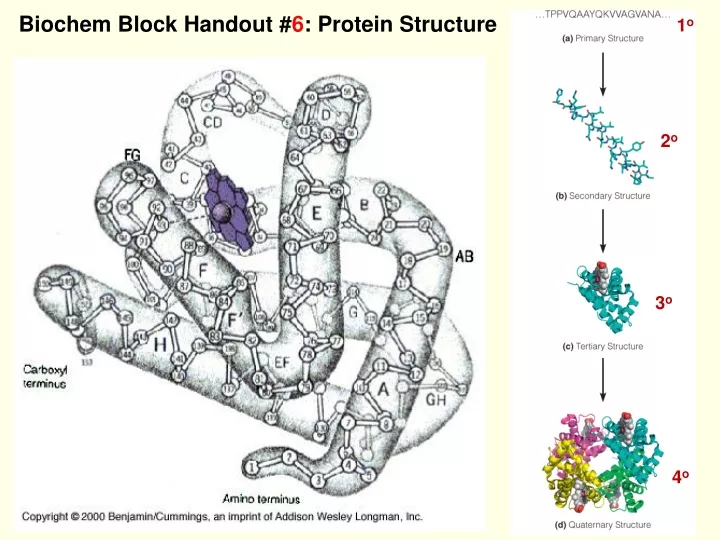
1o 2o Biochem Block Handout #6: Protein Structure 3o 4o
Protein Structure Levels: Definitions • 1º (primary) structure: sequence of aa’s • 2º (secondary) structure: local folding of a polymer to form a regular, repeating structure (local 3-D) • 3º (tertiary) structure: how the 2º structures fit together (global 3-D) • 4º (quaternary) structure: interactions between separate folded polypeptides ex. Hemoglobin • Proteins are held together by H-bonding interactions and other noncovalent interactions
The plane truth about peptide bonds • Both C and N are sp2 hybridized planar • C-N has double bond character can’t rotate • Because of steric constraints, only certain values of(φ,ψ) are allowed
Pitch: (rise) (# residues/turn) Turn: how far one travels to reach a point on the helix directly above the starting point on the helix Repeat: how far one travels along the helical axis in the helix before the structure exactly repeats itself (repeats is an integer value of residues) Repeat = pitch if # residues per turn = an integer Helical Parameters
α-helix • H-bonds between residues separated by 3 aa. C=O of aa #1 bonds with H-N of aa #5, aa #2 with #6, etc. • H-bonds between carbonyl oxygen and hydrogen of amide nitrogen • 3.6 residues per turn • rise (vertical height between residues) = 1.5Ǻ • pitch = (# residues / turn) x (rise) = 5.4 Ǻ. This is the vertical distance of a turn as traveled along the helical axis.
Secondary Structure: Regular Ways to Fold the Polypeptide Chain An antiparallel arrangement of strands forming b-sheets.
Fibrous Proteins: a-keratins Helix filament • Fibrous proteins have a filamentous, or elongated, form. • The a-keratins are made of a-helix and are found in hair, fingernails and skin.
The structure of silk fibroin, a beta sheet protein The b-sheet regions comprise almost exclusively multiple repetitions of the sequence: [Gly-Ala-Gly-Ala-Gly-Ser-Gly-Ala-Ala-Gly-(Ser-Gly-Ala-Gly-Ala-Gly)8] (a) A three-dimensional view of the stacked sheets of fibroin. The region shown contains only alanine and glycine residues. (b) Interdigitation of alanine or serine side chains and glycine side chains in fibroin. The plane of the section is perpendicular to the folded sheets.
Collagen Structure: The basic unit of the collagen fiber is the tropocollagen molecule. A triple helix of three polypeptide chains, ~1000 residues in length. Left-handed helices, with about 3.3 residues/turn. The chains wrap around one another in a right-handed sense. Hydrogen bonds are between the chains, using hydroxyproline and hydroxylysine. Collagen fibers: the material of tendons, bone & skin
Scurvy • Scurvy is a connective tissue disease from a deficiency in Vitamin C (ascorbic acid). • Collagen fibers are weakened. • It is caused by failure to hydroxylate prolines and lysines in collagen. • There is less H-bonding between the chains of tropocollagen.
Post-translational Processing: Some proteins need additional modification to be functional Example: Biosynthesis and assembly of collagen: • Steps 1–4 occur in the endoplasmic reticulum and cytosol of collagen-synthesizing cells. • Steps 6 and 7 occur in the extracellular region.
Globular proteins: • Carry out most of the chemical work of the cell • Synthesis • Transport • Metabolism • Possess secondary structures • Fold into compact tertiary structures • All globular proteins have a defined inside and outside: • hydrophobic residues are packed • mostly on the inside and the hydrophilic residues are on the surface, in contact with water. • Many carry prosthetic groups • small molecules that may be noncovalently or covalently bonded to the protein (e.g., heme in myoglobin). Globular Proteins: Tertiary 3o Structure The tertiary structure of BPTI: • A “stick” model showing atomic positions from X-ray diffraction. • A “cartoon” model of the backbone. • A “surface” model showing the solvent accessible surface.
Turns in 2o structure of proteins b-turns g-turn
Folding, Denaturation & Refolding • Most of the information for determining the 3-D structure of a protein is carried in the amino acid sequence of that protein. • Under harsh conditions, a protein loses its functional 3-D structure. • This process is called denaturation. • Denaturing conditions include: • Increased temperature • pH becomes extremely acidic or alkaline • Organic solvents or urea This schematic drawing depicts the classic RNase A refolding experiment of Anfinsen.
Folding of a globular protein is clearly thermodynamically favorable in vivo • The overall free energy change for folding must be negative. • This negative ΔG is achieved by a balance of several thermodynamic factors: Conformational Entropy Charge–Charge Interactions Internal Hydrogen Bonds van der Waals Interactions The Thermodynamics & Kinetics of Folding Many diseases are associated with protein misfolding: Alzheimer’s, Parkinson’s Mad Cow, ALS, Type II diabetes, etc. Kinetics of folding: some proteins can fold in milliseconds; A random fold of a 125-aa protein takes 1050 years
Quaternary Structure of Proteins Symmetries of protein quaternary structures: Although composed of asymmetric polypeptides, proteins adopt many symmetrical patterns in forming quaternary structures.