Download
1 / 56
950 likes | 1.98k Vues
Antibodies: Structure and Function.

E N D
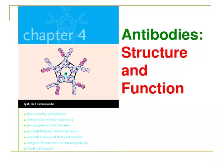
Antibodies: Structure and Function
Antibodiesare antigen-binding proteins present on the B-cell membrane and secreted by plasma cells. Membrane-bound antibody confers antigenic specificity on B cells; antigen-specific proliferation of B-cell clones is elicited by the interaction of membrane antibody with antigen. Secreted antibodies circulate in the blood, where they serve as the effectors of humoral immunity by searching out and neutralizing antigens or marking them for elimination.
1. Basic Structure of Antibodies Experimental demonstration that most antibodies are in the -globulin fraction of serum proteins.
2) Chemical and Enzymatic Methods Revealed Basic Antibody Structure 木瓜蛋白酶 巯基乙醇
2. Obstacles to Antibody Sequencing Initial attempts to determine the amino acid sequence of the heavy and light chains of antibody were hindered because insufficient amounts of homogeneous均一的protein were available. 1) Pure Immunoglobulin Obtained from Multiple Myeloma Patients Made Sequencing Possible
2) Light-Chain Sequencing Revealed That Immunoglobulins Have Constant and Variable Regions
3) Heavy-Chain Sequencing Revealed Five Basic Varieties of Heavy Chains
3. Immunoglobulin Fine Structure The structure of the immunoglobulin molecule is determined by the primary, secondary, tertiary, and quaternary organization of the protein.
1) Immunoglobulins Possess Multiple Domains Based on the Immunoglobulin Fold
2) Diversity in the Variable-Region Domain Is Concentrated in CDRs Because the antigen binding site is complementary to the structure of the epitope, these areas are now more widely called complementarity determining regions (CDRs). The remainder of the VL and VH domains exhibit far less variation; these stretches are called the framework regions (FRs).
3) CDRs Bind Antigen FIGURE 4-10 (a) Side view of the three-dimensional 空间的structure of the combining site of an angiotensin血管紧缩素II–Fab complex. The peptide is in red. The three heavy-chain CDRs (H1, H2, H3) and three lightchain CDRs (L1, L2, L3) are each shown in a different color. All six CDRs contain side chains, shown in yellow, Waals contact of the angiotensin peptide.
(b) Side view of the van der Waals surface of contact between angiotensin II and Fab fragment.
4) Conformational构象的Changes May Be Induced by Antigen Binding Structure of a complex between a peptide derived from HIV protease and an Fab fragment from an anti-protease antibody (left) and comparison of the Fab structure before and after peptide binding (right). In the right panel, the red line shows the structure of the Fab fragment before it binds the peptide and the blue line shows its structure when bound. There are significant conformational changes in the CDRs of the Fab on binding the antigen. These are especially pronounced in the light chain CDR1 (L1) and the heavy chain CDR3 (H3).
5) Constant-Region Domains The immunoglobulin constant-region domains take part in various biological functions that are determined by the amino acid sequence of each domain.
CH1 AND CL DOMAINS-serve to extend the Fab arms of the antibody molecule, thereby facilitating interaction with antigen and increasing the maximum rotation of the Fab arms. HINGE REGION-is rich in proline 脯氨酸residues and is flexible, giving IgG, IgD, and IgA segmental flexibility. As a result, the two Fab arms can assume various angles to each other when antigen is bound. OTHER CONSTANT-REGION DOMAINS IgA, IgD, IgG IgE, IgM CH1/CH1 CH1/CH1 Hinge region CH2/CH2 CH2/CH2 CH3/CH3 CH3/CH3 CH4/CH4
4. Antibody-Mediated Effector Functions In addition to binding antigen, antibodies participate in a broad range of other biological activities. When considering the role of antibody in defending against disease, it is important to remember that antibodies generally do not kill or remove pathogens solely by binding to them. In order to be effective against pathogens, antibodies must not only recognize antigen, but also invoke调用responses—effector functions—that will result in removal of the antigen and death of the pathogen.
3) Antibody-Dependent Cell-Mediated Cytotoxicity (ADCC) Kills Cells
5. Antibody Classes and Biological Activities The various immunoglobulin isotypes同种型;同型;同种and classes have been mentioned briefly already. Each class is distinguished by unique amino acid sequences in the heavy-chain constant region that confer class-specific structural and functional properties. In this section, the structure and effector functions of each class are described in more detail.
1) Immunoglobulin G (IgG) IgG, the most abundant class in serum, constitutes about 80% of the total serum immunoglobulin. The IgG molecule consists of two heavy chains and two or two light chains.
There are four human IgG subclasses, distinguished by differences in -chain sequence and numbered according to their decreasing average serum concentrations: IgG1, IgG2, IgG3, and IgG4.
General structure of the four subclasses of human IgG, which differ in the number and arrangement of the inter chain disulfide bonds (thick black lines) linking the heavy chains. A notable feature of human IgG3 is its 11 interchain disulfide bonds.
The subtle精细的amino acid differences between subclasses of IgG affect the biological activity of the molecule: IgG1, IgG3, and IgG4 readily cross the placenta胎盘and play an important role in protecting the developing fetus. IgG3 is the most effective complement activator, followed by IgG1; IgG2 is less efficient, and IgG4 is notable to activate complement at all. IgG1 and IgG3 bind with high affinity to Fc receptors on phagocytic cells and thus mediate opsonization. IgG4 has an intermediate affinity for Fc receptors, and IgG2 has an extremely low affinity.
2) Immunoglobulin M (IgM) IgM accounts for 5%–10% of the total serum immunoglobulin, with an average serum concentration of 1.5 mg/ml. Monomeric IgM, with a molecular weight of 180,000, is expressed as membrane-bound antibody on B cells. IgM is secreted by plasma cells as a pentamer五聚物in which five monomer units are held together by disulfide bonds that link their carboxyl羧基-terminal heavy chain domains (C4/C4) and their C3/C3 domains
IgM is the first immunoglobulin class produced in a primary response to an antigen, and it is also the first immunoglobulin to be synthesized by the neonate (尤指出生不满一个月的)婴儿. Because of its pentameric由五个部份组成的structure with 10 antigen-binding sites, serum IgM has a higher valency原子价than the other isotypes. An IgM molecule can bind 10 small hapten molecules; however, because of steric原子的)空间(排列)的 位的hindrance, only 5 or fewer molecules of larger antigens can be bound simultaneously. Because of its high valency, pentameric IgM is more efficient than other isotypes in binding antigens with many repeating epitopes such as viral particles and red blood cells (RBCs).
A similar phenomenon occurs with viral particles: less IgM than IgG is required to be required for polymerization聚合作用of the monomers to to neutralize viral infectivity. IgM is also more efficient than IgG at activating complement. Complement activation requires two Fc regions in close proximity, and the pentameric structure of a single molecule of IgM fulfills this requirement.
Because of its large size, IgM does not diffuse well and therefore is found in very low concentrations in the intercellular细胞间的tissue fluids. The presence of the J chain allows IgM to bind to receptors on secretory cells,which transport it across epithelial linings to enter the external secretions that bathe mucosal surfaces. Although IgA is the major isotype found in these secretions, IgM plays an important accessory role as a secretory immunoglobulin.
3) Immunoglobulin A (IgA) Although IgA constitutes only 10%–15% of the total immunoglobulin in serum, it is the predominant . 优越的, 卓越的, 有力的immunoglobulin class in external secretions such as breast milk, saliva唾液, tears, and mucus of the bronchial支气管的, genitourinary泌尿生殖器的, and digestive tracts. In serum, IgA exists primarily as a monomer, but polymeric forms (dimers, trimers, and some tetramers) are sometimes seen, all containing a J-chain polypeptide. The IgA of external secretions, called secretory IgA, consists of a dimer or tetramer, a J-chain polypeptide, and a polypeptide chain called secretory component.
The daily production of secretory IgA is greater than that of any other immunoglobulin class. IgA-secreting plasma cells are concentrated along mucous membrane surfaces. Along the jejunum空肠of the small intestine, for example, there are more than 2.5 X 10¹⁰ IgA-secreting plasma cells—a number that surpasses the total plasma cell population of the bone marrow, lymph, and spleen combined! Every day, a human secretes from 5 g to 15 g of secretory IgA into mucous secretions.
Secretory IgA serves an important effector function at mucous membrane surfaces, which are the main entry sites of any other for most pathogenic organisms. Because it is polymeric, secretory IgA can cross-link large antigens with multiple epitopes. Binding of secretory IgA to bacterial and viral surface antigens prevents attachment of the pathogens to the mucosal cells, thus inhibiting viral infection and bacterial colonization建群. Breast milk contains secretory IgA and many other molecules that help protect the newborn against infection during the first month of life.
4) Immunoglobulin E (IgE) The potent biological activity of IgE allowed it to be identified in serum despite its extremely low average serum concentration (0.3 g/ml). IgE antibodies mediate the immediate hypersensitivity reactions that are responsible for the symptoms of hay fever, asthma哮喘, hives寻麻疹, and anaphylactic shock过敏性休克.
5) Immunoglobulin D (IgD) IgD has a serum concentration of 30 g/ml and constitutes about 0.2% of the total immunoglobulin in serum. IgD, together with IgM, is the major membrane bound immunoglobulin expressed by mature B cells, and its role in the physiology of B cells is under investigation. No biological effector function has been identified for IgD.
6 Antigenic Determinants on Immunoglobulins Since antibodies are glycoproteins, they can themselves function as potent immunogens to induce an antibody response. Such anti-Ig antibodies are powerful tools for the study of B-cell development and humoral immune responses. The antigenic determinants, or epitopes, on immunoglobulin molecules fall into three major categories: isotypic, allotypic[同种]异型, and idiotypic独特型determinants, which are located in characteristic portions of the molecule.
Antigenic determinants of immunoglobulins. For each type of determinant, the general location of determinants within the antibody molecule is shown (left) and two examples are illustrated (center and right). (a) Isotypic determinants are constantregion determinants that distinguish each Ig class and subclass within a species. (b) Allotypic determinants are subtle amino acid differences encoded by different alleles of isotype genes. Allotypic differences can be detected by comparing the same antibody class among different inbred strains. (c) Idiotypic determinants are generated by the conformation of the amino acid sequences of the heavy- and light-chain variable regions specific for each antigen. Each individual determinant is called an idiotope, and the sum of the individual idiotopes is the idiotype.
Monoclonal Antibodies Have Important Clinical Uses Toxins used to prepare immunotoxins include ricin蓖麻毒蛋白, Shigella toxin, and diphtheria toxin白喉毒素. Each toxin contains an inhibitory toxin chain (red) and a binding component (yellow). To make an immunotoxin, the binding component of the toxin is replaced with a monoclonal antibody (blue).