Download
1 / 49
490 likes | 579 Vues
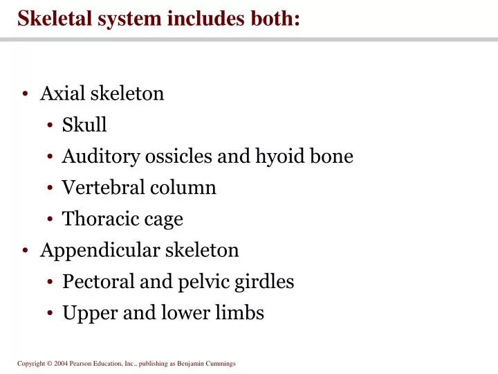
Skeletal system includes both:. Axial skeleton Skull Auditory ossicles and hyoid bone Vertebral column Thoracic cage Appendicular skeleton Pectoral and pelvic girdles Upper and lower limbs. Figure 7.1b The Axial Skeleton. Figure 7.1b. The skull.

E N D
Skeletal system includes both: • Axial skeleton • Skull • Auditory ossicles and hyoid bone • Vertebral column • Thoracic cage • Appendicular skeleton • Pectoral and pelvic girdles • Upper and lower limbs
Figure 7.1b The Axial Skeleton Figure 7.1b
The skull • Consists of the cranium and the bones of the face • The cranium encloses cranial cavity • Facial bones surround and protect the entrances to the respiratory and digestive tracts • Superficial landmarks include the sutures • Lambdoid • Coronal • Sagittal • Squamous
Figure 7.2 Cranial and Facial Subdivisions of the Skull Figure 7.2
Figure 7.3 The Adult Skull Figure 7.3a, b
Figure 7.3 The Adult Skull Figure 7.3c
Figure 7.3 The Adult Skull Figure 7.3d
Figure 7.3 The Adult Skull Figure 7.3e
Focus: The Individual Bones of the Skull Cranial Bones • one occipital bone • foramen magnum • two parietal bones • one frontal bone • frontal sinuses • two temporal bones • auditory ossicles • one sphenoid • one ethmoid
Figure 7.4 The Sectional Anatomy of the Skull Figure 7.4a
Figure 7.4 The Sectional Anatomy of the Skull Figure 7.4b
Figure 7.5 The Occipital and Parietal Bones Figure 7.5
Figure 7.6 The Frontal Bone Figure 7.6
Figure 7.7 The Temporal Bones Figure 7.7
Figure 7.8 The Sphenoid Figure 7.8
Figure 7.9 The Ethmoid Figure 7.9
Facial bones • Inferior nasal conchae • Zygomatic bones • Lacrimal bones • Hyoid • Maxillary bones • Mandible • Palatine bones • Nasal bones • Vomer
Maxillae • Largest facial bones • Form the upper jaw and most of the hard palate
Figure 7.10 The Maxillary and Palatine Bones Figure 7.10
Palatine and Nasal Bones • Palatine bones • Small “L” shaped bones • Form the posterior hard palate and floor of the nasal cavity • Nasal bones • Superior border of external nares
Vomer, Zygomatic and Lacrimal bones • Vomer • Inferior portion of the nasal septum • Zygomatic bone • Temporal process articulates with zygomatic process of temporal bone • Lacrimal bones • Smallest bones of the face • Sit medially in orbit
Figure 7.11 The Smaller Bones of the Face Figure 7.11
Mandible and Hyoid bones • Mandible • Bone of the lower jaw • Hyoid • Suspended by stylohyoid ligaments • Supports the larynx
Figure 7.12 The Mandible and Hyoid Bones Figure 7.12a
Figure 7.12 The Mandible and Hyoid Bones Figure 7.12b, c
The orbital and nasal complexes • Seven bones in the orbital complex • Nasal complex = bones that enclose the nasal cavities and paranasal sinuses
Figure 7.14 The Orbital Complex Figure 7.14
Skulls of infants and children • Fontanels permit skulls of infants and children to continue growing
Vertebral column • Vertebrae, sacrum, coccyx • 7 cervical vertebrae • 12 thoracic vertebrae • 5 lumbar vertebrae • Sacrum and coccyx are fused vertebrae
Figure 7.16 The Vertebral Column Figure 7.16
Spinal curvature • Four spinal curves • Primary (accommodation) curves = thoracic and sacral • Secondary (compensation) curves = lumbar and cervical
Vertebral anatomy • Typically has a body and vertebral arch • Superior and inferior articular processes • Separated by intervertebral discs
Figure 7.18 Vertebral Anatomy Figure 7.18
Vertebral regions • Cervical • Has distinctive shape • Large relative size of vertebral foramen • Costal processes with transverse foramina • Notched spinous processes
Figure 7.19 The Cervical Vertebrae Figure 7.19
Thoracic vertebrae • Heart-shaped body • Long slender spinous processes • Articulations for ribs
Figure 7.20 The Thoracic Vertebrae Figure 7.20a
Figure 7.20 The Thoracic Vertebrae PLAY Animation: Axial Dissections Figure 7.20b, c
Lumbar vertebrae • Most massive • Least mobile • Subjected to great stresses
Figure 7.21 The Lumbar Vertebrae Figure 7.21
Sacrum • Protects reproductive, digestive and urinary organs • Articulates with pelvic girdle and fused elements of coccyx
Figure 7.22 The Sacrum and Coccyx Figure 7.22
Thoracic cage • Thoracic vertebrae • Ribs • Sternum • Ribs and sternum forms the rib cage
Figure 7.23 The Thoracic Cage Figure 7.23a
Figure 7.23 The Thoracic Cage Figure 7.23b
The ribs • Ribs 1-7 are attached to vertebrae • 8-12 are vertebrochondral ribs • 11-12 are floating ribs
Typical rib • Has a head, neck, tubercle and a body • Costal groove marks pathway of blood returning to the heart
The Sternum consists of • Manubrium • Body • Xiphoid process
Figure 7.23 The Thoracic Cage PLAY Animation: Axial Dissections Figure 7.23