Download
1 / 21
220 likes | 409 Vues
Gustation & Vision. Kayla Coggburn, Grace Davis, Carmen Matthews, and Charlie Williams. Gustation is taste Gustation provides information about the foods and liquids that we put into our mouths

E N D
Gustation & Vision Kayla Coggburn, Grace Davis, Carmen Matthews, and Charlie Williams
Gustationis taste • Gustation provides information about the foods and liquids that we put into our mouths • We’re able to taste things because of taste buds, which are sensory structures with specialized epithelial cells • The tongue contains basal cells, which divide in order to produce other cells that mature to become gustatory cells Gustation
Humans have four primary taste sensations: sweet, salty, sour, and bitter • There are also two additional, less common taste sensations: Umami and water • Umami is a pleasant taste that is associated with parmesan cheese and soup broth. Taste Sensations
1. Dissolved chemicals bind to the receptor proteins of the gustatory cells. • 2. Every taste sensation has a different receptor mechanism • 2A. Salty & Sour: Chemically gated ions which result in depolarization • 2B. Sweet, Bitter, and Umami: G proteins called gustducins • 3. Neurotransmitters are released by the receptor cell, resulting in taste receptor stimulation. • 4. The release of the neurotransmitters generates action potential, which leads to taste sensations and reception. Steps to Taste
Humans start life with over 10,000 taste buds, but the number drops around 50. • As people age they lose sensitivity to foods. Foods that young people find spicy, older people find bland and with too little flavor. Aging and Gustation
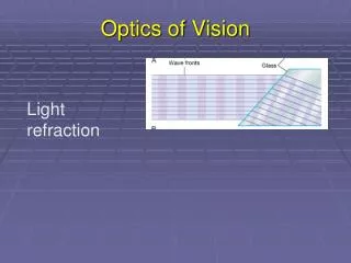
Accessory structures of the eye include: eyelids, superficial epithelium of the eye, and all the structures that are associated with production, secretion, and removal of tears. • Structures that make the exterior of the eye: • Palpebral fissure - Gap that separates free space of the upper and lower eyelids • Medial canthus & lateral canthus – Where the eyelids do connect • Eyelashes – Hairs that prevent objects from entering eye surface • Tarsal glands– Secretes a lipid that prevent eyelids from sticking together • Conjunctiva – Epithelium that covers inner surfaces of the eyelids & outer layer of the eye • Palpebral conjunctiva – Covers inner surface of eyelids • Ocular conjunctiva – Covers anterior surface of the eye • Cornea – A transparent part of the outer layer of the eye Vision-Structures of the Eye
The eyes are an irregular spheroid shape, and weigh about 8 oz. • The eye is cushioned by orbital fat • The eye contains three layers • 1. Outer layer – fibrous tunic • a) Contains the sclera and cornea • 2. Middle layer – vascular tunic • b) Contains blood vessels, lymphatic vessels, and intrinsic muscles • 3. Inner layer – neural tunic (retina) • c) Contains a pigmented part and a neural part The Eye
The retina holds photoreceptors, which are responsible for detecting light • The retina has around 130 million photoreceptors • There are two types of photoreceptors: • 1. Rods – Do NOT determine colors. Allow people to see light • 2. Cones – Color vision • Cones don’t allow you to see color unless there is a strong enough light source to stimulate them • Rods and cones synapse with 6 million bipolar cells, which then synapse with ganglion cells to see color and light • The lens lets our eye focus by changing its shape • An astigmatism may occur if the cornea or lens is curved in an odd way, which causes distortion when looking at something The Retina
Photoreceptors detect photons, which are the basic unit of visible light • The process of detecting and receiving light is called photoreception • Humans are receptive to wavelengths 700-400nm (visible light spectrum, ROY G BIV) • (Red photons = greatest wavelength, least energy; Violet photons = shortest wavelength, most energy) • Rods are able to give the central nervous system the information needed about the presence or absence of photons. (Seeing light) • Cones give the information of the wavelength of photons. (Seeing color) Photoreceptors, Rods & Cones
Sodium ion channels are chemically regulated in the outer portion of the photoreceptor. • In dark: the channels are left open with the presence of cyclic-GMP. Since the channels are open, the transmembrane potential is lower than normal, which makes the photoreceptor releasing neurotransmitters continuously across the inner segment synapses. • That inner layer is simultaneously pumping sodium ions out of the cytoplasm. This is called the dark current. • This is how people are able to vaguely see when the lighting is low Photoreception
Begins when a photon hits a rhodopsin molecule • 4 Steps in rhodopsin-based photoreception: • Step 1– Opsin is activated. • Step 2 – Opsin activates transducin, which then activates phosphodiesterase • Step 3 – Cyclic-GMP levels decline, and the gated sodium channels close • Step 4 – The dark current is reduced, and the rate of neurotransmitters that are released decreases Rhodopsin-based Photoreception
After a photon is absorbed, the whole rhodopsin molecule has to be broken down and the put back together • Bleaching is when a rhodopsin molecule starts to break down into retinene and opsin • Retinene must be enzymatically converted by ATP • Opsin is inactive throughout the process of bleaching • Cyclic-GMP is produced, and the sodium ion channels are reopened Recovery
There are three types of cones: • 1. Blue cones • 16% of all cones • 2. Green cones • 10% of all cones • 3. Red cones • 74% of all cones • Every type of cone has its own form of opsin, and different sensitivities to different wavelength ranges. • The way that the cones are stimulated is the basis for color vision • We see color because of the information received from all three cone types. • Yellow is a combination of highly stimulated green cones, less stimulated red cones, and non-stimulated blue cones • White is a combination of all three cones stimulated equally Color Vision
Color blindness occurs when someone has one or more sets of cones that don’t function • The cones could be absent, or just not able to produce the pigment needed • There is inherited color blindness that involves one to two cones. This is not uncommon. • 10% of men have some form of color blindness, but only 0.67% of women do. • Only 1 person out of 300,000 have total color blindness. Where they have no cones at all. Color Blindness
The visual pathway starts at the photoreceptors and ends at the visual cortex • The message must be sent across two synapses before it can be read. • 1. Photoreceptor to bipolar cell • 2. Bipolar cell to ganglion cell • M cells give information about the general form and shape of an object, its motion, and shadows. • The activation of M cells indicates that light has been detected and has arrived in a general area. • P cells give information about edges, detail and color. Visual Pathway
Astigmatism – A condition that causes blurred vision from one of three reasons: • Irregular shape of the cornea • Irregular shape of the cover of the eye • Curvature of the lens inside the eye • Conjunctivitis – An inflammation of the conjunctiva (transparent layer of tissue that surrounds the inner part of the eye and the white part) Otherwise known as “pink eye”. It can be caused by: • Allergies • Bacteria • Cataract – A cloudy, or yellow, part of the clear and transparent lens of the eye. Can be caused by: • Diabetes • Smoking • Certain Drugs • Nutritional deficiency Diseases
Ophthalmologist - A medical doctor who specializes in eye and vision care • Optometrist – A healthcare professional who provides vision care. Things like sight testing and correction, diagnosis of the problem, treatments, and manages changes in vision. • Optician – A technician who designs, verifies and fits glasses lenses, frames and contact lenses Careers