Download
1 / 84
850 likes | 1k Vues
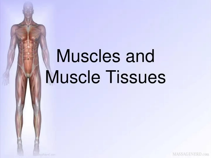
Muscles and Muscle Tissues. Overview. Types of Muscles Tissues Skeletal Cardiac Smooth These types differ in structure, location, function, and means of activation. Similarities. Skeletal and smooth muscle cells are elongated and called muscle fibers

E N D
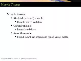
Overview • Types of Muscles Tissues • Skeletal • Cardiac • Smooth • These types differ in structure, location, function, and means of activation
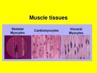
Similarities • Skeletal and smooth muscle cells are elongated and called muscle fibers • Muscle contractions depend on two types of myofilaments • Actin • Myosin • Muscle terminology is similar • Sarcolemma – muscle plasma membrane • Sarcoplasm – cytoplasm of a muscle cell • Prefixes – myo, mys, and sarco all refer to muscle
Skeletal Muscle Tissues • Packaged in Skeletal Muscles that attach to and cover the bony skeleton • Has obvious stripes called • Striations • Controlled voluntarily • Contracts rapidly, but tires easily • Is responsible for overall body motility • Is extremely adaptable and can exert forces over a range from a fraction of an ounce to over 70 pounds
Cardiac Muscle Tissue • Occurs only in the Heart • Striated like Skeletal Muscle but not Striated • Contracts at a • fairly steady rate by the heart’s pacemaker • Neutral Control allows heart to respond to changes in bodily needs
Smooth Muscle Tissue • Found in the walls of hollow visceral organs, such as the • Stomach • Urinary bladder • Respiratory Passages • Forces food and other substances through internal body channels • It is not striated and is involuntary
Muscle Function • Skeletal muscles are responsible for all locomotion • Cardiac muscle is responsible for coursing the blood through the body • Smooth muscle helps maintain blood pressure, and squeezes or propels substances (i.e., food, feces) through organs • Muscles also • maintain posture • stabilize joints • generate heat
Functional Characteristics of Muscles • Excitability, or irritability – the ability to receive and respond to stimuli • Contractility – the ability to shorten forcibly • Extensibility – the ability to be stretched or extended • Elasticity – the ability to recoil and resume the original resting length
Skeletal Muscles • Each muscle is a discrete organ composed of muscle tissue, blood vessels, nerve fibers, and connective tissue • Three Connective Tissue Wrappings • Epimysium – an overcoat of dense regular CT that surrounds the entire muscle • Perimysium – fibrous CT that surrounds groups of muscle fibers called fascicles • Endomysium – fine sheath of CT composed of reticular fibers surrounding each muscle fiber
Skeletal Muscle:Nerve and Blood Supply • Each muscle is served by one nerve, an artery, and one or more veins • Each skeletal muscle fiber is supplied with a nerve ending that controls contraction • Contracting fibers require continuous delivery of oxygen and nutrients via arteries • Wastes must be removed via veins
Skeletal Muscle: Attachments • Muscles span joints and are attached to bone in at least two places • When muscles contract the movable bone, the muscle’s insertion moves toward the immovable bone – the muscle’s origin • Muscles attach: • Directly – epimysium of the muscle is fused to the periosteum of a bone • Indirectly – CT wrappings extend beyond the muscle as a tendon or aponeurosis
Microscopic Anatomy of a Skeletal Muscle Fiber • Each fiber is a long, cylindrical cell with multiple nuclei just beneath the sarcolemma • Each fiber is 10 to 100 m in diameter and up to hundreds of centimeters long • Each cell is a syncytium produced by fusion of embryonic cells
Microscopic Anatomy of a Skeletal Muscle Fiber • Sarcoplasm has numerous glycosomes and a unique oxygen-binding protein called myoglobin • Fibers contain • organelles • myofibrils • sarcoplasmic reticulum • T tubules
Myofibrils • Myofibrils are densely packed, rodlike contractile elements • They make up most of the muscle volume • The arrangement of myofibrils within a fiber is such that a perfectly aligned repeating series of dark A bands and light I bands is evident
Sarcomeres • The smallest contractile unit of a muscle • The region of a myofibril between two successive Z discs • Composed of myofilaments made up of contractile proteins • Myofilaments are of two types • Thick • Thing
Myofilaments:Banding Patterns • Thick filaments – extend the entire length of an A band • Thin filaments – extend across the I band and partway into the A band • Z-disc – coin-shaped sheet of proteins (connectins) that anchors the thin filaments and connects myofibrils to one another • Thin filaments do not overlap thick filaments in the lighter H zone • M lines appear darker due to the presence of the protein desmin
Ultrastructure of Myofilaments:Thick Filaments • Composed of the protein myosin • Each myosin molecule has a rodlike tail and two globular heads • Tails – two interwoven, heavy polypeptide chains • Heads – two smaller, light polypeptide chains called cross bridges
Ultrastructure of Myofilaments:Thin Filaments • Thin filaments are chiefly composed of the protein actin • Each actin molecule is a helical polymer of globular subunits called G actin • The subunits contain the active sites to which myosin heads attach during contraction • Tropomyosin and troponin are regulatory subunits bound to actin
Arrangement of Filaments in a Sacromere • Longitudinal section within one sarcomere
Sarcoplasmic Reticulum (SR) • SR is an elaborate smooth endoplasmic reticulum that mostly runs longitudinally and surrounds each myofibril • Paired terminal cisternae form perpendicular cross channels • Functions in the regulation of intracellular calcium levels • Elongated tubes called T tubules penetrate into the cell’s interior at each A band–I band junction • T tubules associate with the paired terminal cisternae to form triads
T Tubles • T tubules are continuous with the sarcolemma • They conduct impulses to the deepest regions of the muscle • These impulses signal for the release of Ca2+ from adjacent terminal cisternae
Contraction of Skeletal Muscle Fibers • Contraction – refers to the activation of myosin’s cross bridges (force generating sites) • Shortening occurs when the tension generated by the cross bridge exceeds forces opposing shortening • Contraction ends when cross bridges become inactive, the tension generated declines, and relaxation is induced
Sliding Filament Method of Contraction • Thin filaments slide past the thick ones so that the actin and myosin filaments overlap to a greater degree • In the relaxed state, thin and thick filaments overlap only slightly • Upon stimulation, myosin heads bind to actin and sliding begins • Each myosin head binds and detaches several times during contraction, acting like a ratchet to generate tension and propel the thin filaments to the center of the sarcomere • As this event occurs throughout the sarcomeres, the muscle shortens
Role of Ionic Calcium (Ca2+) in the Contraction Mechanism • At low intracellular Ca2+ concentration: • Tropomyosin blocks the binding sites on actin • Myosin cross bridges cannot attach to binding sites on actin • The relaxed state of the muscle is enforced
Role of Ionic Calcium (Ca2+) in the Contraction Mechanism • At higher intracellular Ca2+ concentrations: • Additional calcium binds to troponin (inactive troponin binds two Ca2+) • Calcium-activated troponin binds an additional two Ca2+ at a separate regulatory site
Role of Ionic Calcium (Ca2+) in the Contraction Mechanism • Calcium-activated troponin undergoes a conformational change • This change moves tropomyosin away from actin’s binding sites • Myosin head can now bind and cycle • This permits contraction (sliding of the thin filaments by the myosin cross bridges) to begin
Sequential Events of Contraction • Cross bridge attachment – myosin cross bridge attaches to actin filament • Working (power) stroke – myosin head pivots and pulls actin filament toward M line • Cross bridge detachment – ATP attaches to myosin head and the cross bridge detaches • “Cocking” of the myosin head – energy from hydrolysis of ATP cocks the myosin head into the high energy state
Regulation of Contraction • In order to contract, a skeletal muscle must: • Be stimulated by a nerve ending • Propagate an electrical current, or action potential, along its sarcolemma • Have a rise in intracellular Ca2+ levels, the final trigger for contraction • Linking the electrical signal to the contraction is excitation-contraction coupling
Nerve Stimulus of Skeletal Muscle • Skeletal muscles are stimulated by motor neurons of the somatic nervous system • Axons of motor neurons branch profusely as they enter muscles • Each axonal branch forms a neuromuscular junction with a single muscle fiber
Neuromuscular Junction • The neuromuscular junction is formed from: • Axonal endings, which have small membranous sacs (synaptic vesicles) that contain the neurotransmitter acetylcholine (ACh) • The motor end plate of a muscle, which is a specific part of the sarcolemma that contains ACh receptors that helps form the neuromuscular junction • Though exceedingly close, axonal ends and muscle fibers are always separated by a space called the synaptic cleft
Neuromuscular Junction • When a nerve impulse reaches the end of an axon at the neuromuscular junction: • Voltage-regulated calcium channels open and allow Ca2+ to enter the axon • Ca2+ inside the axon terminal causes axonal vesicles to fuse with the axonal membrane • This fusion releases ACh into the synaptic cleft via exocytosis • ACh diffuses across the synaptic cleft to ACh receptors on the sarcolemma • Binding of ACh to its receptors initiates an action potential in the muscle
Action Potential • A transient depolarization event that includes polarity reversal of a sarcolemma (or nerve cell membrane) and the propagation of an action potential along the membrane
Action Potential: Electrical Conditions of a Polarized Sarcolemma • The outside (extracellular) face is positive, while the inside face is negative • This difference in charge is the resting membrane potential • The predominant extracellular ion is Na+ • The predominant intracellular ion is K+ • The sarcolemma is relatively impermeable to both ions
Action Potential: Depolarization and Generation of the Action Potential • An axonal terminal of a motor neuron releases ACh and causes a patch of the sarcolemma to become permeable to Na+ (sodium channels open) • Na+ enters the cell, and the resting potential is decreased (depolarization occurs) • If the stimulus is strong enough, an action potential is initiated
Action Potential: Propagation of the Action Potential • Polarity reversal of the initial patch of sarcolemma changes the permeability of the adjacent patch • Voltage-regulated Na+ channels now open in the adjacent patch causing it to depolarize • Thus, the action potential travels rapidly along the sarcolemma • Once initiated, the action potential is unstoppable, and ultimately results in the contraction of a muscle
Action Potential: Repolarization • Immediately after the depolarization wave passes, the sarcolemma permeability changes • Na+ channels close and K+ channels open • K+ diffuses from the cell, restoring the electrical polarity of the sarcolemma • Repolarization occurs in the same direction as depolarization, and must occur before the muscle can be stimulated again (refractory period) • The ionic concentration of the resting state is restored by the Na+-K+ pump
Destruction of Acetylcholine • ACh bound to ACh receptors is quickly destroyed by the enzyme acetylcholinesterase (AChE) • AChE activity prevents continued muscle fiber contraction in the absence of additional stimuli
Excitation-Contraction Coupling • Once generated, the action potential: • Is propagated along the sarcolemma • Travels down the T tubules • Triggers Ca2+ release from terminal cisternae • Ca2+ binds to troponin and causes: • The blocking action of tropomyosin to cease • Actin active binding sites to be exposed • Myosin cross bridges alternately attach and detach • Thin filaments move toward the center of the sarcomere • Hydrolysis of ATP powers this cycling process • Ca2+ is removed into the SR, tropomyosin blockage is restored, and the muscle fiber relaxes
Contraction of Skeletal Muscle (Organ Level) • Contraction of muscle fibers (cells) and muscles (organs) is similar • The two types of muscle contractions are: • Isometric contraction – increasing muscle tension (muscle does not shorten) • Isotonic contraction – decreasing muscle length (muscle shortens during contraction)
Motor Unit: The Nerve-Muscle Functional Unit • A motor unit is a motor neuron and all the muscle fibers it supplies • The number of muscle fibers per motor unit can vary from four to several hundred • Muscles that control fine movements (fingers, eyes) have small motor units • Large weight-bearing muscles (thighs, hips) have large motor units • Muscle fibers from a motor unit are spread throughout the muscle; therefore, contraction of a single motor unit causes weak contraction of the entire muscle
Muscle Twitch • A muscle twitch is the response of a muscle to a single brief threshold stimulus • The three phases of a muscle twitch are: • Latent period – first few milliseconds after stimulation when excitation-contraction coupling is taking place • Period of contraction – cross bridges actively form and the muscle shortens • Period of relaxation – Ca2+ is reabsorbed into the SR, and muscle tension goes to zero
Graded Muscle Response • Graded muscle responses are: • Variations in the degree of muscle contraction • Required for proper control of skeletal movement • Responses are graded by: • Changing the frequency of stimulation • Changing the strength of the stimulus
Muscle Response to Varying Stimuli • A single stimulus results in a single contractile response – a muscle twitch • Frequently delivered stimuli (muscle does not have time to completely relax) increases contractile force – wave summation • More rapidly delivered stimuli result in incomplete tetanus • If stimuli are given quickly enough, complete tetanus results
Muscle Response: Stimulation Strength • Threshold stimulus – the stimulus strength at which the first observable muscle contraction occurs • Beyond threshold, muscle contracts more vigorously as stimulus strength is increased • Force of contraction is precisely controlled by multiple motor unit summation • This phenomenon, called recruitment, brings more and more muscle fibers into play
Treppe: The Staircase Effect • Staircase – increased contraction in response to multiple stimuli of the same strength • Contractions increase because: • There is increasing availability of Ca2+ in the sarcoplasm • Muscle enzyme systems become more efficient because heat is increased as muscle contracts