Download
1 / 52
520 likes | 607 Vues
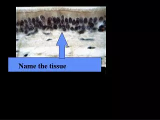
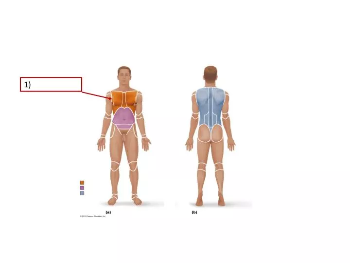
1). 2). Name this tissue (Remove the box for a hint). 3). Name this tissue (Remove the box for a hint). 4). Name this tissue (Remove the box for a hint). 5). 7). 6). 8). 10). 9). 11). Name this Joint. 12).

E N D
7) 6)
10) 9)
Name this Joint 12)
Name this Joint(you can move the boxes for more examples of this kind of joint) 13)
Name the Joint (again, you can move the boxes to see more examples) 14)
15) 16)
20) 19)
21) 23) 22) 24)
25) 26)
27) 29) 28)
30) 32) 31)
33) 34) 35)
36) 37)
38) 39) 40)
41) 42) 43)
47) 45) 46)
48) 49)
Figure 16.2 Cyclic AMP second-messenger mechanism of water-soluble hormones. 50) 51)
Figure 16.3 Direct gene activation mechanism of lipid-soluble hormones. 52) 53)
54) 55)
56) 57)
58) 59)
60) 61)
64) 65)
66) 67)
69) 70)
Figure 17.2 Photomicrograph of a human blood smear stained with Wright’s stain. Baroreceptor reflexes that help maintain blood pressure homeostasis 71) 72)
Figure 20.2a The lymphatic system. 75) 76) 74) 78) 77)
Figure 22.8 Respiratory zone structures. 81) Name this single grapelike structure:82)
Figure 23.1 Alimentary canal and related accessory digestive organs. 84) 85) 86)
Figure 25.1 The urinary system. 89) 90) Region where arteries, veins enter the kidney? 92) 91)
93) 94) 95)
66) 97) 98)
Answer Key • 19. Primary Motor Cortex • 20. Primary Somatosensory Cortex • 21. Corpus Callosum • 22. Septum Pellucidum • 23. Choroid Plexus • 24. Arbor Vitae • 25. Oculomotor Nerve • 26. Vestibulocochlear Nerve • 27. Gray Commissure • 28. Arachnoid Mater • 29. Lateral Funiculus • 30. Ciliary Body • 31. Retina • 32. Macula Lutea • 33. Superior Oblique • 34. Inferior Rectus • 35. Inferior Oblique • 1. Axillary2. Superior mediastinum (in the thoracic cavity)3. Fibrocartilage Connective Tissue • 4. Smooth muscle connective tissue • 5. Stratified Squamous Epithelium6. Radius • 7. Olecranon Process • 8. Xiphoid Process • 9. Acetabulum • 10. Superior Ramus of Pubis • 11. Tibial Tuberosity • 12. Diarthrotic synovial condyloid • 13. Synarthrotic Fibrous Joint • 14. Amphiarthrotic Cartilaginous joint • 15. Latissimusdorsi • 16. Teres Major • 17. Pectineus • 18. Zygomaticus bicep