Download
1 / 41
420 likes | 582 Vues
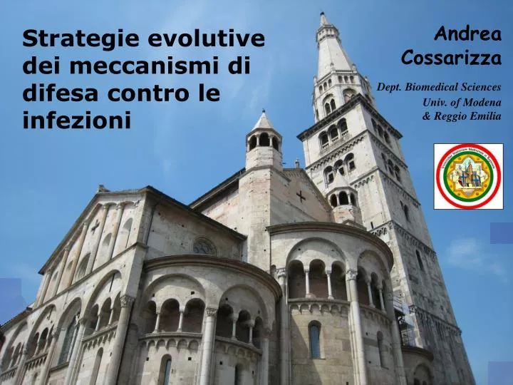
Andrea Cossarizza. Strategie evolutive dei meccanismi di difesa contro le infezioni. Dept. Biomedical Sciences Univ. of Modena & Reggio Emilia. MAIN TOPICS. NKT cells - distribution and characteristics - biological role. MAIN TOPICS. NKT cells - distribution and characteristics

E N D
Andrea Cossarizza Strategie evolutive dei meccanismi di difesa contro le infezioni Dept. Biomedical Sciences Univ. of Modena & Reggio Emilia
MAIN TOPICS NKT cells - distribution and characteristics - biological role
MAIN TOPICS NKT cells - distribution and characteristics - biological role ANTIGEN PRESENTATION by CD1 - molecules and distribution - functional role
MAIN TOPICS NKT cells - distribution and characteristics - biological role ANTIGEN PRESENTATION by CD1 - molecules and distribution - functional role TOLL LIKE RECEPTORS - ligands and role - evolutionary aspects - counteractions by pathogens
MAIN TOPICS NKT cells - distribution and characteristics - biological role ANTIGEN PRESENTATION by CD1 - molecules and distribution - functional role TOLL LIKE RECEPTORS - ligands and role - evolutionary aspects - counteractions by pathogens
INNATE IMMUNE SYSTEM Because of its critical role in initiating and orchestrating a productive immune response, it is imperative to obtain a detailed understanding of the microbial determinants that are recognized by different components and cells of the innate immune system. One member of the innate immune system, the natural killer T (NKT) cell, is activated during bacterial infections.
NATURAL KILLER T CELL (NKT) • NKT cells constitute a unique subpopulation of T lymphocytes that is highly conserved, and well evident in both human and murine species. • The term 'NKT cells' was first used to describe a small subset of the T lymphocytes that coexpressed some markers traditionally associated with NK cells. • The most prominent of these markers was the NK1.1 antigen (NRK-P1/CD161 in humans), which has regularly been used in conjunction with TCR expression to phenotypically identify NKT cells. • At first glance, NKT cells may be easily mistaken as being part of the adaptive immune system.
T CELL RECEPTOR EXPRESSION Like conventional T cells, NKT cells express T-cell receptors that are generated via somatic DNA rearrangement.
T CELL RECEPTOR EXPRESSION The TCRs of most NKT cells are semi-invariant, consisting of Vα14–Jα18/Vβ8.2 chains in mouse and homologous Vα24 – Jα18 / Vβ11 chains in human (iNKT cells).
NATURAL KILLER T CELL • In humans, only 0.2%ofperipheral blood T cells are NKT cells • They are also present in the human liver and play a crucial role in the control of cells infected by hepatitis virus • NKT cells express markers associated with recently activated or memory T cells
CO-RECEPTOR EXPRESSION CD4 • Human and mouse NKT cells segregate into CD4+CD8− and CD4−CD8− (double negative, DN) cell subsets, which differ in their functional properties. • In humans, about 40 to 60% of invariant NKT cells are CD4+, with high donor-to-donor variability, whereas the remaining cells lack CD4 expression. • In humans, CD8α expression is common, but only very few CD8β+ NKT cells exist (CD8 expression is even absent in mice) • The CD4+ subset potently produces both Th1 and Th2 cytokines • DN population selectively produces the Th1 cytokines (IFN-γ) and TNF-a and preferentially upregulates perforin in response to IL-2 or IL-12 CD8
ANTIGENS RECOGNITION: CD1d • NKT cells are activated by glycolipid antigens presented in the monomorphic MHC I-like molecule CD1d highly conserved among mammalian species.
CD1 MOLECULES CD1 molecules comprise a family of glycoproteins that resemble class I MHC molecules in their domain organization. Humans possess one of each of the five CD1 isoforms, located on chromosome 1, in the order shown. Mice and rats appear to have duplicated CD1d and lost the genes for CD1a, CD1b, CD1c and CD1e, possibly in a chromosomal translocation event.
Ag presentation by CD1 The discovery of molecules capable of presenting lipid antigens and of the T cells that recognize them has opened a new dimension in our understanding of cell-mediated immunity against infection. Like MHC Class I molecules, CD1 isoforms (CD1a, b, c and d) are assembled in the ER and sent to the cell surface. However, in contrast to MHC molecules, CD1 complexes are then re-internalized into specific endocytic compartments where they can bind lipid antigens.
Assembly of nascent CD1 molecules in ER is assisted by chaperones (orange). Microsomal triglyceride transfer protein (MTP, blue) assists CD1 loading with ER-resident self-GSL (red). In LE/Ly, LTP (light blue) extract lipids from membranes and participate in loading and unloading of CD1 molecules. Soluble CD1e is involved in processing of large microbial glycolipids (in green) by binding and offering them to hydrolases (yellow). Upon internalization of bacteria (green cells), microbial lipids are released into phagosomes and loaded onto CD1 molecules. Infection, by altering self-lipid metabolism facilitates CD1–self-lipid complex generation and activation of autoreactive T cells. De Libero et al. Eur J Imm, Oct. 09
Assembly of nascent CD1 molecules in ER is assisted by chaperones (orange). Microsomal triglyceride transfer protein (MTP, blue) assists CD1 loading with ER-resident self-GSL (red). In LE/Ly, LTP (light blue) extract lipids from membranes and participate in loading and unloading of CD1 molecules. Soluble CD1e is involved in processing of large microbial glycolipids (in green) by binding and offering them to hydrolases (yellow). Upon internalization of bacteria (green cells), microbial lipids are released into phagosomes and loaded onto CD1 molecules. Infection, by altering self-lipid metabolism facilitates CD1–self-lipid complex generation and activation of autoreactive T cells. De Libero et al. Eur J Imm, Oct. 09
Assembly of nascent CD1 molecules in ER is assisted by chaperones (orange). Microsomal triglyceride transfer protein (MTP, blue) assists CD1 loading with ER-resident self-GSL (red). In LE/Ly, LTP (light blue) extract lipids from membranes and participate in loading and unloading of CD1 molecules. Soluble CD1e is involved in processing of large microbial glycolipids (in green) by binding and offering them to hydrolases (yellow). Upon internalization of bacteria (green cells), microbial lipids are released into phagosomes and loaded onto CD1 molecules. Infection, by altering self-lipid metabolism, facilitates the generation of CD1–self-lipid complex and the consequent activation of autoreactive T cells. De Libero et al. Eur J Imm, Oct. 09
Ag presentation by CD1 CD1 molecules are differentially expressed by a variety of cell types including DC, B cells, monocytes, Langerhans cells, stellate hepatic cells, epithelial cells, microglial cells and keratinocytes. CD1a, CD1b, CD1c and CD1d molecules reach the plasma membrane and are involved in lipid presentation to T cells, whereas CD1e remains intracellular and is involved in lipid processing. Lipid-specific T cells express a variety of TCR heterodimers, with the exception of invariant NKT (iNKT) cells. Lipid-loaded CD1 molecules delivered to the cell surface are surveyed by CD1-restricted T cells expressing ab or gd TCR.
CD1 PRESENT A WIDE VARIETY OF LIPIDS Endogenous cellular lipids, foreign lipids derived from intracellular parasites and extracellular lipids of self or foreign origin. The degree to which different CD1 isoforms are specialized for binding structurally distinct lipids remains unclear CD1a present a mycobacterial lipopeptide in addition to glycolipids CD1b accommodates lipids with very long alkyl chains (e.g. C80; mycolates that are components of mycobacterial cell walls) CD1c present mycobacterial isoprenoid lipids that contain unusual alkyl chains consisting of repeating branched, unsaturated units. Some lipids, including phospholipids and sphingolipids, have been shown to bind multiple CD1 isoforms including CD1a, CD1b, CD1c and CD1d
Structures of self-lipid antigens. iGb3: isoglobotrihexosylceramide; PG: phosphatidylglycerol; PE: phosphatidylethanolamine; PI: phosphatidylinositol.
Structures of microbial lipid antigens. a-galactosylceramide (a-GalCer) extracted from A. mauritianus. BbGL-II: Borrelia burgdorferi diacylglycerol; GMM: glucose monomycolate; GroMM: glycerol monomycolate.
iNKT cell activation During infection, iNKT cells are rapidly elicited. Activated iNKT cells can produce a vast array of cytokines (IFN-γ, IL-4, IL-2, IL-5, IL-10, IL-13, GM-CSF, TNF-α …...) that affect the innate and the adaptive immune response
Ag presentation by CD1hot points: The capacity of the immune system to recognize lipid antigens relies on a series of biochemical and biological characteristics of lipid molecules. These are associated with the structure of lipids, which affects their bioavailability, type of trafficking and capacity to associate with CD1 molecules. It also depends on the structure of CD1 molecules that have evolved different antigen-binding pockets and trafficking capacity.
Ag presentation by CD1hot points: An important difference between MHC and CD1 antigen presenting molecules is that CD1 are functionally non-polymorphic and this results from selective pressure dictated by the structure of microbial lipid antigens. While peptides lose their MHC-binding capacity by changing single amino acids, lipids are much more constrained in changing their CD1-binding moieties because these modifications are often not compatible with their biological function.
MAIN TOPICS NKT cells - distribution and characteristics - biological role ANTIGEN PRESENTATION by CD1 - molecules and distribution - functional role TOLL LIKE RECEPTORS - ligands and role - evolutionary aspects - counteractions by pathogens
A family of transmembrane receptors that have been highly preserved throughout evolution. Toll-like receptors (TLRs)
Neighbour-joining molecular tree of the extracellular domains of mammalian TLRs
Sequence evolution in the extracellular domain after gene duplication might be the key to understanding how the ligand binding affinity evolves. Positive pressure Purifying selection w = dN/ds (non-synonymous/ synonymous substitution ratio) Analysis of a dataset of 22 mammalian TLR2 sequences (black line), compared to sequences of ten ruminant TLR2 sequences (red line)
A family of transmembrane receptors that have been highly preserved throughout evolution.They selectively recognize a broad spectrum of microbial components (PAMPs: Pathogen Associated Molecular Pattern) and endogenous molecules released by injured tissue Toll-like receptors (TLRs)
ANTI-IMMUNE EVOLUTION OF PATHOGENSAnti-interferon mechanisms
Activation of the interferon response triggered by the detection of viral PAMPs
Activation of the interferon response triggered by the detection of viral PAMPs
Activation of the interferon response triggered by the detection of viral PAMPs
Viral evasion of retinoic-acid-inducible-gene-(RIG) i-like receptor signalling
Inhibition of interferon-regulatory factor 3 (iRF3) and iRF7 by viral proteins
CONCLUSIONS • DURING EVOLUTION, THE IMMUNE SYSTEM HAS DEVELOPED SEVERAL STRATEGIES TO FIGHT INFECTIONS; • INFECTIVE AGENTS HAVE DEVELOPED SEVERAL STRATEGIES TO COPE WITH THE IMMUNE SYSTEM. • THE GENERAL SURGEON WAS NOT RIGHT…… THERE WILL BE ALWAYS SOMETHING TO DO – INCLUDING BOOKS TO WRITE - FOR US!
Thanks for your attention