Download
1 / 42
520 likes | 1.47k Vues
Coagulation Disorders and Disseminated Intravascular Coagulation (DIC). Jianzhong Sheng MD, PhD Department of Pathophysiology School of Medicine Zhejiang University. Normal Hemostasis. First step in hemostasis is formation of a platelet aggregate

E N D
Coagulation Disorders and Disseminated Intravascular Coagulation (DIC) Jianzhong Sheng MD, PhD Department of Pathophysiology School of Medicine Zhejiang University
Normal Hemostasis • First step in hemostasis is formation of a platelet aggregate • At the molecular level interaction of coagulation factors takes place on the surface of activated platelets • The Tissue Factor–FVIIa complex is the physiological activator of normal hemostasis
Hemostasis Endothelial cell Subendothelial matrix Nitric oxide
Coagulation Pathways Intrinsic Pathway Extrinsic Pathway Tissue Factor + VII IX TF Pathway Contact X TF-VIIa XI Common Pathway PL XIIa HKa Prothrombin XIa PL (Tenase) IXa PL VIIIa Xa XIII Va (Prothrombinase) Thrombin Protein C, Protein S, Antithrombin III XIIIa Fibrinogen Fibrin (weak) Fibrin (strong)
Xa VIIa TF Va TF-Bearing Cell Normal Hemostasis X II IIa (Thrombin)
TF VIIa Xa Va TF-Bearing Cell Normal Hemostasis II X VIII/vWF IIa VIIIa
TF VIIa Xa Va TF-Bearing Cell Normal Hemostasis II X VIII/vWF IIa VIIIa V Va Platelet
TF VIIa Xa Va TF-Bearing Cell Normal Hemostasis II X VIII/vWF IIa VIIIa V Va Platelet Activated Platelet
TF VIIa Xa Va TF-Bearing Cell Normal Hemostasis II X VIII/vWF IIa VIIIa TF V Va VIIa IX Platelet IXa Activated Platelet
TF VIIa Xa Va TF-Bearing Cell Normal Hemostasis II X VIII/vWF IIa VIIIa TF V Va VIIa IX Platelet II IXa X Xa IIa VIIIa IXa Va Activated Platelet
Normal Hemostasis II X VIII/vWF TF VIIa Xa IIa Va VIIIa TF-Bearing Cell TF V Va VIIa IX Platelet II IXa X Xa IIa VIIIa IXa Va Activated Platelet Hoffman et al. Blood Coagul Fibrinolysis 1998;9(suppl 1):S61.
Normal Hemostasis: Pivotal role of TF/VIIa II X VIII/vWF VIIa TF Xa IIa Va VIIIa TF-Bearing Cell TF V Va VIIa IX Platelet II IXa X Xa IIa VIIIa IXa Va Activated Platelet VIIa IXa Va IIa Xa VIIIa II IX X Hoffman et al. Blood Coagul Fibrinolysis 1998;9(suppl 1):S61.
ADP Aggregation Aggregation Aggregation GpIIb/IIIa GpIIb/IIIa GpIIb/IIIa GpIIb/IIIa GpIIb/IIIa GpIIb/IIIa Adrenaline Adhesion Adhesion vWF Endothelium Exposed Collagen Platelet Activation Pathways COLLAGEN THROMBIN ADP Hemophilia GpIIb/IIIa Platelet GpIb Adrenaline Adhesion Gp Glycoprotein
Bleeding through a Cut in a Vessel Wall Tissue Factor Factor VIIa • The first step in all coagulation: The Tissue Factor- Factor VIIa complex formation
Bleeding through a Cut in a Vessel Wall TissueFactor- Factor VIIa Complex • The first step in all coagulation: The Tissue Factor- • Factor VIIa complex formation • This catalysis the coagulation cascade in normal persons and in patients with bleeding disorders
Recombinant Factor VIIa Platelet Binding TissueFactor- Factor VIIa Complex rFactorVIIa Platelets Recombinant Factor VIIa (rFVIIa) in high concentration binds to platelets; this complex catalysis further coagulation. The local coagulation activation is greatly enhanced
Further Formation of a Hemostatic Plug TissueFactor-rFVIIa Complex rFVIIa Platelets High peak levels of recombinant Factor VIIa (rFVIIa) induces formation of a strong fibrin network. This network cross-binds and forms a solid hemostatic plug
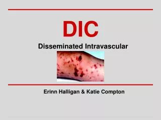
Primarily a thrombotic process Systemic process producing both thrombosis and hemorrhage Also called consumption coagulopathy and defibrination syndrome1 Its clinical manifestation may be widespread hemorrhage in acute, fulminant cases2. -Background -Pathophysiology -Etiology -Clinical Manifestations -Diagnosis -Treatment -Xigris DIC
Basic pathophysiology Entry into the circulation of procoagulant substances Trigger systemic activation of the coagulation system and platelets Lead to the disseminated deposition of fibrin-platelet thrombi. Procoagulant stimulus is tissue factor (most cases) Lipoprotein Not normally exposed to blood. Tissue factor gains access to blood by Tissue injury, Malignant cells, Expression on the surfaces of monocytes and endothelial cells by inflammatory mediators. -Background -Pathophysiology -Etiology -Clinical Manifestations -Diagnosis -Treatment DIC
Tissue factor triggers Thrombin Protease Induces fibrin formation and platelet activation Other procoagulants Cysteine protease Mucin(粘液素) Trypsin -Background -Pathophysiology -Etiology -Clinical Manifestations -Diagnosis -Treatment DIC
Acute DIC Coagulation factors are consumed at a rate in excess of the capacity of the liver to synthesize them, Platelets are consumed in excess of the capacity of bone marrow megakaryocytes to release them. -Background -Pathophysiology -Etiology -Clinical Manifestations -Diagnosis -Treatment DIC
Laboratory manifestations Prolonged prothrombin time (PT) Prolonged Activated partial thromboplastin time (aPTT) Thrombocytopenia. Increased fibrin formation Stimulates compensatory process of secondary fibrinolysis, Plasminogen activators generate plasmin to digest fibrin (and fibrinogen) into fibrin(ogen) degradation products (FDPs). FDPs are potent circulating anticoagulants that contribute further to the bleeding manifestations of DIC. Intravascular fibrin deposition can cause fragmentation of red blood cells and lead to the appearance of schistocytes in blood smears Hemolytic anemia is unusual in DIC. Microvascular thrombosis in DIC can compromise the blood supply to some organs and lead to multiorgan failure -Background -Pathophysiology -Etiology -Clinical Manifestations -Diagnosis -Treatment DIC
DIC • -Background • -Pathophysiology • -Etiology • -Clinical Manifestations • -Diagnosis • -Treatment
DIC always has an underlying etiology Must be identified and eliminated to treat the coagulopathy(凝血病) successfully. The development of DIC in many of these disorders is associated with an unfavorable outcome1. Occurs in 1% of hospitalized patients Mortality rate approaches 40-80% -Background -Pathophysiology -Etiology -Clinical Manifestations -Diagnosis -Treatment DIC
Causes Infection Most common cause of DIC. The syndrome particularly is associated with gram-negative or gram-positive sepsis Can be triggered by a variety of other Bacterial Fungal Viral Rickettsial, and protozoal microorganisms. -Background -Pathophysiology -Etiology -Clinical Manifestations -Diagnosis -Treatment DIC
Obstetrics The placenta and uterine contents are rich sources of Tissue factor Other procoagulants that normally are excluded from the maternal circulation -Background -Pathophysiology -Etiology -Clinical Manifestations -Diagnosis -Treatment DIC
Clinical manifestations of DIC may accompany obstetric complications, especially in the third trimester. These syndromes range from Acute, fulminant, and often fatal DIC in amniotic fluid embolism Blood is exposed to large amounts of tissue factor in a short period of time creating large amounts of thrombin Multiorgan failure Chronic or subacute DIC with a retained dead fetus. Exposure to small amounts of tissue factor -Background -Pathophysiology -Etiology -Clinical Manifestations -Diagnosis -Treatment DIC
Other obstetric problems associated with DIC include Abruptio placentae(胎盘剥离) Toxemia Septic abortion. -Background -Pathophysiology -Etiology -Clinical Manifestations -Diagnosis -Treatment DIC
Clinical manifestations Determined by Nature Intensity Duration of the underlying stimulus. Chronicity Low-grade DIC is often asymptomatic Diagnosed only by laboratory abnormalities. Bleeding is most common clinical finding Generalized or widespread ecchymoses Chronic disease Thrombotic complications Trousseau's syndrome in cancer Gangrene of the digits or extremities Hemorrhagic necrosis of the skin Purpura fulminans Enhanced by Coexistence of liver disease -Background -Pathophysiology -Etiology -Clinical Manifestations -Diagnosis -Treatment DIC
Diagnosis of severe, acute (easy) Prolongation of PT, aPTT and Thrombin time Due to consumption and inhibition of clotting factors Thrombocytopenia Fibrin degradatin products Increased due to secondary fibrinolysis Measured by latex agglutination or D-dimer assays. Schistocytes may be seen in the peripheral blood smear Neither sensitive nor specific for DIC. -Background -Pathophysiology -Etiology -Clinical Manifestations -Diagnosis -Treatment DIC
Chronic or compensated forms of DIC Highly variable patterns of abnormalities in "DIC screen" coagulation tests. Increased FDPs and prolonged PT are generally more sensitive measures than are abnormalities of the aPTT and platelet count. Overcompensated synthesis of consumed clotting factors and platelets in some chronic forms Cause shortening of the PT and aPTT and/or thrombocytosis Though, elevated levels of FDPs indicate secondary fibrinolysis in such cases. -Background -Pathophysiology -Etiology -Clinical Manifestations -Diagnosis -Treatment DIC
Treatment Identify underlying cause and treat All other therapies are temporizing -Background -Pathophysiology -Etiology -Clinical Manifestations -Diagnosis -Treatment DIC
Asymptomatic patients with self-limited DIC Have only laboratory manifestations of the coagulopathy No treatment may be necessary. -Background -Pathophysiology -Etiology -Clinical Manifestations -Diagnosis -Treatment DIC
Actively bleeding or who are at high risk of bleeding, Blood component treatments of choice Transfusions of platelets Improve the thrombocytopenia Fresh-frozen plasma (FFP) Replace all consumed coagulation factors and correct the prolonged PT and aPTT. Large volumes of plasma in severe cases The theoretical concern that these blood products may "fuel the fire" and exacerbate the DIC has not been supported by clinical experience -Background -Pathophysiology -Etiology -Clinical Manifestations -Diagnosis -Treatment DIC
Special cases Profound hypofibrinogenemia Additional transfusion of cryoprecipitate, Plasma concentrate enriched in fibrinogen Sepsis Infusion of antithrombin III concentrate may be considered as an adjunctive measure -Background -Pathophysiology -Etiology -Clinical Manifestations -Diagnosis -Treatment DIC
Pharmacologic inhibitors of coagulation and fibrinolysis Heparin Theoretical benefit It blocks thrombin and the secondary fibrinolysis. Might exacerbate the bleeding tendency Usually reserved for Forms manifested by Thrombosis Acrocyanosis (发绀) Cancer Vascular malformations Retained dead fetus Acute promyelocytic leukemia. 早幼粒细胞 -Background -Pathophysiology -Etiology -Clinical Manifestations -Diagnosis -Treatment DIC
Antifibrinolytic agents, ε-aminocaproic acid and tranexamic acid Generally are contraindicated May precipitate thrombosis May be effective in decreasing life-threatening bleeding -Background -Pathophysiology -Etiology -Clinical Manifestations -Diagnosis -Treatment DIC