Download
1 / 24
261 likes | 1.27k Vues
Objectives. Assess the anatomy, physiology, and pathophysiology of the tonsillectomy

E N D
1. Tonsillectomy & Adenoidectomy See Exemplar Provided
2. Objectives Assess the anatomy, physiology, and pathophysiology of the tonsillectomy & adenoidectomy
Analyze the diagnostic and surgical interventions for a patient undergoing a _______________.
Plan the intraoperative course for a patient undergoing_____________.
Assemble supplies, equipment, and instrumentation needed for the procedure.
3. Objectives Choose the appropriate patient position
Identify the incision used for the procedure
Analyze the procedural steps for_____________.
Describe the care of the specimen
Discuss the postoperative considerations for a patient undergoing _______________
4. Terms & Definitions A
5. Definition/Purpose of Procedure Removal of tonsils & adenoids by sharp or blunt dissection
Adenoids are removed to facilitate breathing, prevent recurrent otitis media, and to restore hearing loss due to obstruction of the eustachian tube
6. Relevant A & P Pharynx (throat) p. 618 � 620 Begins at internal nares and ends posterior to the larynx at the level of the esophagus. It is divided into 3 regions according to location: the nasopharynx, oropharynx, and laryngopharynx. The lymphoid elements (pharngeal, palatine, and lingual tonsils, and the pharngeal band) contained with the pharynx are named Waldeyer�s ring.
What is the difference between the palatine tonsils and the lingual tonsils? What are the pharyngeal tonsils?
Palatine: (also called faucial)�two oval masses of lymphoid tissue which are located at each edge of the fauces withing the folds of two bands of tissue that descend from the soft palate to the base of the tongue, called the tonsillar pillars. Each tonsil has an anterior and posterior pillar. Behind each posterior posterior tonsillar pillar is a pink band of lymphoid tissue called the lateral pharyngeal band. The tonsils produce lymphocytes. Each of the palantine tonsils contains 10-20 crypts or dips that help trap bacteria.
The lingual tonsils are a pair of lymphoid areas located on the posterior surface of the tongue near the base.
The pharyngeal tonsils are are located on the posterior wall of the nasopharynx. When the pharyngeal tonsils are enlarged, they are referred to as adenoids. They are a single mass of lymphatic tissue embedded n the mucous membrane of the nasopharynx. These tonsils help to protect against pathogens entering the nose. After about age 7, this lymphatic tissue usually begins to shrink.
Pharynx (throat) p. 618 � 620 Begins at internal nares and ends posterior to the larynx at the level of the esophagus. It is divided into 3 regions according to location: the nasopharynx, oropharynx, and laryngopharynx. The lymphoid elements (pharngeal, palatine, and lingual tonsils, and the pharngeal band) contained with the pharynx are named Waldeyer�s ring.
What is the difference between the palatine tonsils and the lingual tonsils? What are the pharyngeal tonsils?
Palatine: (also called faucial)�two oval masses of lymphoid tissue which are located at each edge of the fauces withing the folds of two bands of tissue that descend from the soft palate to the base of the tongue, called the tonsillar pillars. Each tonsil has an anterior and posterior pillar. Behind each posterior posterior tonsillar pillar is a pink band of lymphoid tissue called the lateral pharyngeal band. The tonsils produce lymphocytes. Each of the palantine tonsils contains 10-20 crypts or dips that help trap bacteria.
The lingual tonsils are a pair of lymphoid areas located on the posterior surface of the tongue near the base.
The pharyngeal tonsils are are located on the posterior wall of the nasopharynx. When the pharyngeal tonsils are enlarged, they are referred to as adenoids. They are a single mass of lymphatic tissue embedded n the mucous membrane of the nasopharynx. These tonsils help to protect against pathogens entering the nose. After about age 7, this lymphatic tissue usually begins to shrink.
7. Relevant A & P See Video clip www.nucleusinc.com
8. Pathophysiology Upper aerodigestive tract
Tonsillitis of the palatine tonsils STST p. 619, -622.
Located in the Oropharynx
Common preop diagnosis is �tonsillar hypertrophy� or �Recurrent tonsillitis.�
Other problems which can occur in that region are peritonsillar abscess and cancer.
Tonsillitis may affect any or all of the tonsillar regions�but it is usually of the palatine tonsils. The problem can be acute or chronic. Usually it�s caused by strepotococcal organism.
Acute:enlarged and dusky and purulence of tonsillar crypts.
Chronic: persistent sore throat, foul breat, enlarged cervical lymph nodes.
Failure to treat chronic tonsillitis can lead to a peritonsillar abscess�or if treated and antibiotics failed. (Fig 17-28 p. 622) S & S: extreme pain, difficulty breathing, referred pain to the ear on the affected side. Surgery is called an I & D (incision and drainage). This is an emergency due to potential airway obstruction AND the infection can spread quickly to neck and chest, causing complications like pericarditis (fatal).
Adenoiditis�inflammation of pharyngeal tonsils�usually bacterial, but may be viral or due to allergies. Recurrent adenoiditis can lead to hypertrophy (snoring is a symptom) or hearing impairment due to eustachian tube blockage.
STST p. 619, -622.
Located in the Oropharynx
Common preop diagnosis is �tonsillar hypertrophy� or �Recurrent tonsillitis.�
Other problems which can occur in that region are peritonsillar abscess and cancer.
Tonsillitis may affect any or all of the tonsillar regions�but it is usually of the palatine tonsils. The problem can be acute or chronic. Usually it�s caused by strepotococcal organism.
Acute:enlarged and dusky and purulence of tonsillar crypts.
Chronic: persistent sore throat, foul breat, enlarged cervical lymph nodes.
Failure to treat chronic tonsillitis can lead to a peritonsillar abscess�or if treated and antibiotics failed. (Fig 17-28 p. 622) S & S: extreme pain, difficulty breathing, referred pain to the ear on the affected side. Surgery is called an I & D (incision and drainage). This is an emergency due to potential airway obstruction AND the infection can spread quickly to neck and chest, causing complications like pericarditis (fatal).
Adenoiditis�inflammation of pharyngeal tonsils�usually bacterial, but may be viral or due to allergies. Recurrent adenoiditis can lead to hypertrophy (snoring is a symptom) or hearing impairment due to eustachian tube blockage.
9. Terms & Definitions Hypertrophy Hypertrophy is enlargement of a structure due to the increase in the size of the cellsHypertrophy is enlargement of a structure due to the increase in the size of the cells
10. Diagnostics Exams
H & P
Visual exam
C & S
Preop testing
CBC: PTT-7 minutes Preoperative testing which should be reported for the patient undergoing tonsillectomy would be PTT-7 minutes. This would be abnormal and indicates bleeding problems. This should be reported to the surgeon because one of the major complications of tonsillectomy is postop bleeding. Preoperative testing which should be reported for the patient undergoing tonsillectomy would be PTT-7 minutes. This would be abnormal and indicates bleeding problems. This should be reported to the surgeon because one of the major complications of tonsillectomy is postop bleeding.
11. Surgical Intervention:Special Considerations OR table position
Order of extraction varies
Best technique (not sterile)
Surgeon may prefer to stand or sit
Typical peds
Adults: under local and sitting up
12. Surgical Intervention: Anesthesia General
Peds mask induction
Oral ET tube
Lubricate and protect eyes
13. Surgical Intervention: Positioning Supine, neck hyperextended
Supplies and equipment
Neck roll
Arm sleds or draw sheet
Safety strap
Foam headrest or donut
Move patient to edge for ease of access
Special considerations: high risk areas
14. Surgical Intervention: Skin Prep N/A
15. Surgical Intervention: Draping/Incision Head wrap or cover sheet
Peritonsillar incision
16. Surgical Intervention: Supplies General
� sheet x 2; raytex; towels; gowns & gloves, suction tubing, ESU w/extension; small basin
Specific
Suture: 2-0 plain heavy, tapered 5/8 in needle
Meds: local of choice (marcaine or lidocaine w/epinephrine)
Tonsil sponges
# 12 knife blade
Insulated suction cautery
Syringe for irrigation
Supplies for local (needle, syringe)
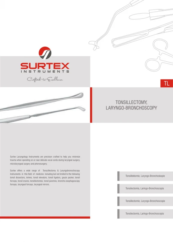
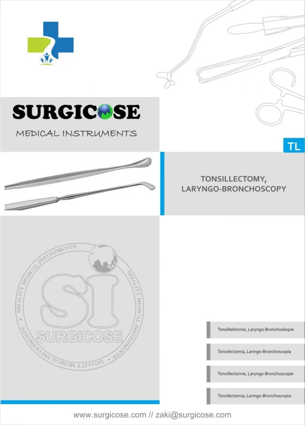
17. Surgical Intervention: Instruments T & A set
18. Surgical Intervention: Equipment Sitting stool
Headlamp
? Harmonic scalpel
Suction apparatus
19. Surgical Intervention: Procedure Steps See Exemplar and Procedure 17-6 STST
20. Counts Initial: sponges and sharps
First closing
Final closing
Sponges
Sharps
21. Specimen & Care Rt and left tonsils and adenoids
Ask about separating�may �tag rt w/safety pin�
22. Postoperative Care Destination PACU�outpatient
Position pt on side once extubated
Elevate HOB
Cold fluids
Expected prognosis
Return to normal activities within 2 wks
Reduced incident of sore throat & ear infections
23. Postop Care Complications
Hemorrhage up to 10 days post op
Infection
Wound Classification : II�increased for inflammation or infection
24. Resources STST Procedure 17-6
STST pp. 618