Download
1 / 26

E N D
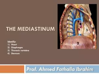
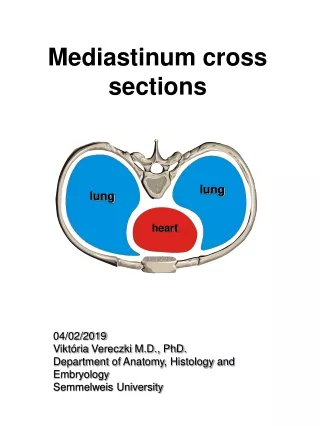
Mediastinum • The mediastinum is the central compartment of the thoracic cavity. It is covered on each side by mediastinal pleura and contains all the thoracic viscera and structures except the lungs. It extends from superior thoracic aperture to the diaphragm inferiorly and from the sternum and costal cartilages anteriorly to bodies of thoracic vertebra posteriorly.
Divisions of Mediastinum • Mediastinum is artificially divided into superior and inferior parts for purposes of description. Superior Mediastinum extends inferiorly from superior thoracic aperture to the horizontal plane which includes the sternal angle anteriorly and junction of T4 and T5 vertebrae posteriorly. Inferior Mediastinum is further subdivided into anterior,middle and posterior parts.
Posterior Mediastinum • RelationsAnteriorly: Pericardium and DiaphragmPosteriorly: T5-T12 VertebraeOn both sides: Parietal pleura of lungsContents:Thoracic Aorta, Azygous and Hemiazygous veins, Posterior Mediastinal Lymph Nodes, Thoracic Duct,Thoracic Sympathetic Trunk, Esophagus, Esophageal Plexus, Thoracic Splanchnic Nerves.
Thoracic Aorta • The thoracic part of descending aorta is the continuation of Arch of Aorta.
Course • It begins on the left side of inferior border of T4 vertebra and descends in the posterior mediastinum on the left sides of T5 to T12 vertebra. As it descends, it approaches the median plane and displaces the esophagus to the right.
Continued • The thoracic aortic plexus, an autonomic nerve network, surrounds it. The thoracic aorta lies posterior to the root of left lung, the pericardium and esophagus. The thoracic aorta terminates(changes into abdominal aorta) anterior to T12 vertebra and enters the abdomen through the aortic opening in the diaphragm. Thoracic Duct and Azygous vein accompany Aorta through this opening.
Relations • Anteriorly: root of the left lung, the pericardium, the esophagus, and the diaphragm. • Posteriorly: vertebral column and the azygos vein • On the right side: with the hemiazygos veins and thoracic duct • On the left side, with the left pleura and lung.
The esophagus, with its accompanying plexus of nerves, lies on the right side of the aorta above; but at the lower part of the thorax it is placed in front of the aorta, and, close to the diaphragm, is situated on its left side.
Branches • Broncial • Pericardial • Posterior Intercostal • Superior Phrenic • Esophageal • Mediastinal • Subcostal
The Bronchial arteries consist of one right and two left vessels. They supply trachea, bronchi, lung tissue, and lymph nodes. • The pericardial arteries supply pericardium. • Posterior Intercostal arteries supply 3rd to 11th Intercostal spaces. • Superior phrenic arteries pass to the posterior surface of diaphraghm, where they anastamose with musculophrenic and pericardiophrenic branches of Internal Thoracic artery.
Two esophageal arteries supply the middle one-third of the esophagus. • Mediastinal arteries are small and supply lymph nodes and other tissues in the posterior mediastinum. • Subcostal arteries enter the abdomen.
Esophagus • The Esophagus descends into the posterior mediastinum from the superior mediastinum, passing posterior to the arch of aorta, pericardium and left atrium. It deviates to the left and passes through the diaphragm via Esophageal opening, at the level of T10 vertebra(accompanied by Vagus Nerves,Branches of Lt Gastric vessels).
Esophageal Impressions/Constrictions • Esophagus is compressed by three structures. • Aortic Arch • Left Main Bronchus • Diaphragm
Esophagus is a tubular structure about 10 inches(25 cm) long that starts opposite C6 vertebra, and is continuous above with the laryngeal portion of pharynx. Relations of thoracic part of Esophagus are • Anteriorly: Trachea,left recurrent laryngeal nerve(RLN), Left Main Bronchus, Pericardium. • Posteriorly: Thoracic Vertebrae, Thoracic Duct, Azygous Veins, Posterior I/C arteries • Right Side: Mediastinal Pleura • Left Side: Left Subclavian artery, Aortic Arch, Thoracic Duct.
Blood Supply • Upper 1/3rd: Inferior Thyroid Artery. Venous Drainage by Inferior thyroid veins • Middle 1/3rd: Descending Thoracic Aorta,drainage by azygous veins • Lower 1/3rd: Left Gastric Artery.Drainage by left gastric vein.
Lymphatic Drainage • From upper 1/3rd: Deep Cervical Nodes • Middle 1/3rd: Superior and Posterior Mediastinal nodes • Lower 1/3rd: Along the left gastric vessels and celiac nodes
Nerve Supply • Supplied by Parasympathetic and sympathetic fibers from vagi and sympathetic trunks.
Clinical Importance • The constrictions are of considerable clinical importance because they are sites where swallowed foreign bodies can lodge or through which endoscope cannot easily pass.