Download
1 / 22

E N D
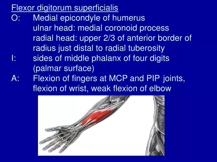
Flexor digitorum superficialisO: Medial epicondyle of humerus ulnar head: medial coronoid process radial head: upper 2/3 of anterior border of radius just distal to radial tuberosityI: sides of middle phalanx of four digits (palmar surface)A: Flexion of fingers at MCP and PIP joints, flexion of wrist, weak flexion of elbow
Intermediate Layer (Anterior) Flexor digitorum superficialis
Movements of the Thumb • Flexion and Extension • Abduction and Adduction
Helpful Hints • “pollicis” – thumb (1st) • “indicis” – index finger (2nd) • “pollicis” and “indicis” exclusive to wrist and fingers
Flexor digitorum profundusO: Proximal ¾ anterior and medial ulnaI: Base of distal phalanx of four fingersA: Flexion of four fingers at MCP, PIP, and DIP joints, wrist flexion
Flexor pollicis longusO: Middle anterior surface of radius and anterior medial border of ulna distal to coronoid processI: Base of distal phalanx of thumb (palmar surface)A: flexion of thumb CMC, MCP, IP joints, flexion, abduction of wrist
Deep Layer (Anterior) • Flexor digitorum profundus • Flexor pollicis longus • *Pronator Quadratus
Extensor indicisO: Middle to distal 1/3 of posterior ulnaI: Base of middle and distal phalanxes of 2nd phalange (dorsal surface)A: Extension of index finger at MCP joint, weak wrist extension, weak supination of forearm from pronated position
Extensor pollicis longusO: Posterior lateral surface of lower middle ulnaI: Base of distal phalanx of thumb (dorsal surface)A: Extension of thumb at CMC, MCP, and IP joints, wrist extension and abduction, weak supination from a pronated position
Extensor pollicis brevisO: Posterior surface lower middle radiusI: Base of proximal phalanx of thumb (dorsal surface)A: Extension of thumb at CMC and MCP joint, weak wrist extension and abduction
Abductor pollicis longusO: Posterior aspect of radius and midshaft of ulnaI: Base of 1st metacarpal (dorsal surface)A: Abduction and extension of thumb at CMC joint, abduction of wrist, weak supination of forearm from a pronated position
Deep Layer (Posterior) • Extensor indicis • Extensor pollicis longus • Extensor pollicis brevis • Abductor pollicis longus • *Supinator
Static Equilibrium • Whenever an object is completely motionless, it is in static equilibrium • For an object to be in static equilibrium: 1) Fv = 0 2) Fh = 0 3) T = 0
Example: How much force must be exerted by the biceps brachii, attaching at 90º to the radius at 3 cm from the elbow joint, to support a weight of 70 N held in the hand, a distance of 30 cm from the elbow joint?
Known: dm = 3 cm = 0.03 m dwt = 30 cm = 0.3 m wt = 70 N Fm = ?
T = 0 T = (Fm)(dm) - (wt)(dwt) = 0
Example: How much force must be exerted by the biceps brachii, attaching at 100º to the radius at 3 cm from the elbow joint, to support a weight of 70 N held in the hand, a distance of 30 cm from the elbow joint?
Carpal Tunnel Syndrome • Swelling of tissue within the compartment causes compression on the median nerve • Symptoms include: • Pain and numbness along the median nerve • Clumsiness of finger function • Weakness and atrophy of muscles • Most common in middle-aged women or older