Download
1 / 32
330 likes | 606 Vues
Cummings Ch 115: Penetrating and Blunt Trauma to the Neck. Kimanh Nguyen May 29, 2013. Vital Structures. Air passages Trachea, larynx, pharynx, lungs Vascular Carotid, jugular, subclavian , innominate , aortic arch Gastrointestinal Pharynx, esophagus Neurologic

E N D
Cummings Ch 115:Penetrating and Blunt Trauma to the Neck Kimanh Nguyen May 29, 2013
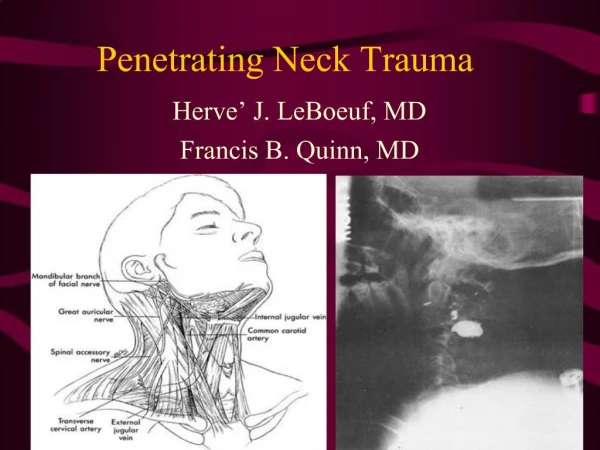
Vital Structures • Air passages • Trachea, larynx, pharynx, lungs • Vascular • Carotid, jugular, subclavian, innominate, aortic arch • Gastrointestinal • Pharynx, esophagus • Neurologic • Spinal cord, brachial plexus, peripheral nerves, cranial nerves
Kinetic Energy • Kinetic energy affects magnitude of injury: • KE = ½ M (V1 – V2)2
Handguns • Projectile type • Speed • Handguns/pistols are low velocity (90-600 m/s) • Caliber • .44-caliber magnum is comparable to a rifle • Yaw • Tumbling bullet causes injury in a wider path
Rifles • Military bullets • Jacket creates smoother flight, clean hole, through-and-through wound • High velocity (760 m/s) transmits energy waves to surrounding tissue • Hunting rifles with expanding bullets • Soft-tips expand, create large wound cavity, may not exit, may fragment • High mortality
Shotguns • Velocity ~ 300 m/s • Distance • Pellets scatter at longer distances • Type of weapon • Sawed-off shotgun sprays the shot earlier • Size of projectile (shot) • Birdshots (< 3.5 mm, 12m range) • Buckshots (> 3.5 mm, 150m range). Comparable to handgun bullet wounds • Wadding
Stab Injuries • Single-entry vs multiple stab wounds • Higher incidence of subclavian vessel laceration due to downward direction • Lower incidence of spinal injuries
Immediate surgical exploration • Massive bleeding • Expanding hematoma • Nonexpanding hematoma with hemodynamic instability • Hemomediastinum • Hemothorax • Hypovolemic shock
Management • “For the stable patient, the choice of management remains controversial: either mandatory exploration for all penetrating neck wounds or selective exploration with observation [and monitoring]”
Zone I • Vascular structures are in close proximity to thorax • Protection by bony thorax and clavicle • Difficult to explore • Median sternotomy for R injuries • Left anterior thoracotomy for L injuries • High mortality rate: 12% • Management: • Angiography if stable • Mandatory exploration usually not recommended • May consider barium swallow
Zone III • Protected by skeletal structures • Difficult to explore; may need craniotomy for high carotid injury • CN injuries may indicate great vessel injury • Management • Angiography if abnormal neurologic exam in stable patient • Frequent intraoral examination for edema/hematoma
Zone II • Most common region injured (60-75%) • Isolated venous and pharyngoesophageal injuries are most commonly missed • Management • Admit for observation • Radiology and endoscopy if stable and no signs of major injury
Initial Management • Airway establishment • Intubation • Cricothyroidotomy • Tracheostomy • Blood perfusion maintenance • Large-bore IV • Clarification and classification of wound severity • Do not probe wound • Routine AP/lat neck and chest films
Vascular Penetration • Zone I • Thoracic surgery • Zone III • Temporary pressure or carotid arterial bypass • No. 4 Fogarty catheter • Jugular • Ligation • Carotid • Ligation of ECA • Lateral arteriorhaphy, end-to-end anastomosis, autogenous grafting • IR transcatheter arterial embolization
Digestive Tract Injury • Gastrograffin swallow • Barium swallow • Flexible esophagoscopy (risk of missing perforations near CP and hypopharynx) • Rigid esophagoscopy • Neck exploration for subQ emphysema or mediastinitis; localization with methylene blue • Management of esophageal injury • 2-layer closure with wound irrigation, debridement, drainage, possible muscle flap • Lateral cervical esophagostomy, later definitive repair
Laryngotracheal Injury • Repair mucosal lacerations within 24 hours • Soft laryngeal stent for badly macerated mucosa • 6-week trach below or through the injury for significant injuries that detach a tracheal ring or encroach on the airway
Blunt Neck Injury • Occult cervical spine injury • Delayed onset of signs and symptoms • Careful observation • Thrombosis, intimal tears, dissection, pseudoaneurysm
Neck Masses • History (time course, risk factors, symptoms) • Physical exam (full head and neck exam, flexible laryngoscopy) • Imaging
Initial workup • Antibiotic trial • Further investigation for concerning signs/symptoms • Unilateral, enlarging, asymmetric, supraclavicularfossae, not associated with infections • Imaging • Biopsy • FNA (gold standard), repeat FNA, core needle biopsy, open biopsy, neck dissection (SCCA)
Inflammatory Neck Masses • Lymphadenopathy/lymphadenitis • Staph, Strep, HIV, lymphoma • Granulomatous disease • TB, MAI, actinomycosis, cat-scratch, syphilis • Sialadenitis/sialolithiasis • Purulent material expressed from ducts
Congenital Neck Masses • Rule out malignancy in adults • Thyroglossal duct cyst • Midline neck mass that elevates with tongue protrusion or swallowing • Rule out median ectopic thyroid • Sistrunk procedure • Branchial cleft anomalies • Cyst, sinus, or fistula • 1st arch (1%), 2nd arch (95%), 3rd and 4th arch (rare) • Complete excision of the tract
Congenital Neck Masses • Dermoid cyst • Trapped rests of epithelial elements • Ectoderm and endoderm • Teratomas • Ectoderm, mesoderm, endoderm • Lymphangiomas • Soft, compressible, 50% present at birth • Hemangiomas • Soft, compressible, bluish-purple, thrill/bruit, 50% regress by age 5
Primary Neoplasms of the Neck • Lymphoma • Most common H&N malignancy in children • 80% of HL have cervical disease • 33% of NHL have cervical disease (90% B-cell) • Thyroid neoplasms • 90% of thyroid nodules are benign • Salivary gland neoplasms • 80% parotid, 15% SMG
Primary Neoplasms of the Neck • Neurogenicneoplasms • Schwannoma (most common), neurofibromas, malignant peripheral nerve sheath tumors, neuromas • Paragangliomas • Neuroectoderm origin, secrete catecholamines • Carotid body, jugulotympanic region, vagus nerve • 10% autosomal dominant/syndromic, 10% multicentric, <10% malignant • Salt and pepper appearance on T1-MRI • Lipomas • Mostly in posterior neck
Unknown Primary SCCA • Thorough physical exam • Imaging of the head, neck, and chest • Panendoscopy and biopsies (BOT, tonsils, NP, HP)