Download
1 / 19
190 likes | 414 Vues
Catalytic strategies. What are the sources of catalytic power and selectivity of enzymes? We investigate here a specific class of enzymes: Ser-proteases . Mechanism of action of these enzymes got determined by a great selection of state-of-the-art biochemical and biophysical approaches

E N D
Catalytic strategies What are the sources of catalytic power and selectivity of enzymes? We investigate here a specific class of enzymes: Ser-proteases. Mechanism of action of these enzymes got determined by a great selection of state-of-the-art biochemical and biophysical approaches including structure determination or site-directed mutagenesis. In each class of Es, comparison between class members reveal how Es and their ASs have evolved and been refined to deliver specific tasks. Struc- tural and mechanistic comparisons of E actions provide us with insight to evolution of Es. These insights lead to design more potent drugs.
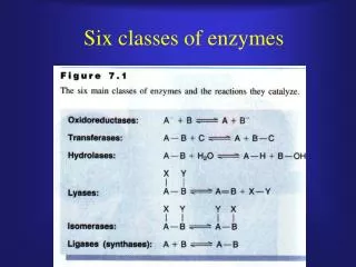
Types of catalytic strategies: Covalent catalysis: AS contains a reactive group, often a nucleophile, that temporarily covalently binds to S. Acid-base catalysis: a H+ donor or acceptor plays role, e.g. in chymotrypsin a His is a base catalyst to enhance the nucleophilicity of Ser. Catalysis by approximation: based simply on binding the two Ss in a single binding surface. Metal ion catalysis: Metal ions can act several ways, e.g. make nucleophiles like OH- by direct coordination (like Zn2+ in carbonic anhydrase), serve as electrophile stabilizing a – charge in an intermediate (like Mg2+ in EcoRV, an endonuclease), serve as bridge between E and S (holding S in place for catalysis and increase binding energy, e.g. in NMP kinases and almost all enzymes that use ATP as S).
Proteases Important in protein degradation and turnover, and in specific regulatory processing of selected enzymes and proteins. Mechanism: hydrolysis by adding H2O to the peptide bond (although thermodinamically favored, it would be very slow without E [10-103 years]). The resonance structure (partial double-bond character) makes peptide bonds planar and kinetically stable. The C of C=O gets protected against nucleophilic attack. The E should facilitate this nucleophilic attack.
Irreversible inactivation occurs. Chymotrypsin Cleaves the the C-terminus of bulkier, hydrophobic AAs, like Trp, Tyr, Phe, Met. Good example of a covalent catalyst. It applies a powerful nucleophile (Nu) to attack the unreactive carbonyl C. The Nu briefly gets covalently attach- ed to the S during the course of catalysis. What is that powerful Nu in chymotrypsin? The clue came from the reac- tion with DIPF that showed one extraordinarily reactive Ser among the 28 Ser residues in the E.
(pre-steady state) They used a chromogenic ester as S (proteases often can hydrolyze esters, too) to monitor E activity. The reaction obeys M-M kinetics with a KM of 20 mM and a kcat of 77 s-1. They used the stopped-flow technique that rapidly mixes E and S (in 1-2 ms) and is able to detect changes spectrophotometrically right after this fast mixing. This proves that hydrolysis proceeds in 2 steps: the burst phase is because the 1st step is faster than the 2nd one. yellow color
The 2 steps are explained by the formation of a covalent E-S intermediate. fast slow (intermediate) p-NO2-phenol The 3D structure of chymotrypsin (solved in 1967) shows 3 polypeptide chains linked by S-Ss. It is synthesized as a single polypeptide (chymo- trysinogen) which is activated by proteolysis that provides the 3 chains.
The active site S catalytic triad general base catalyst (strong Nu) Roles: His positions Ser and polarize its –OH that it would get deprotonated easier. Asp orients His and makes it a better H+-acceptor through H-bonding and electrostatic effects. These observations suggest a mechanism for peptide hydrolysis:
protein N-Hs stabilize the negative charge and the TS S (unstable) mixture of acid-base and covalent catalysis
The mechanism does not account for the cleavage preference of the E. 3D structure with inhibitors and S analogs revealed a deep hydrophobic pocket, called S1 pocket, into which the long, uncharged side-chains of e.g. Phe or Trp can just fit. Binding an appropriate side chain into the pocket positions the adjacent peptide bond into the AS for cleavage.
Cas of trypsin and chymotrypsin are superimposed, rmsd=1.7 Å Other proteases have more complex specificity patterns: Similar ASs and mechanisms are found in homolog Es (e.g. trypsin, elastase). There is ~40% sequence identity and very similar 3D structures among the- se enzymes. However, their S specificities are very different; trypsin cleaves after Lys or Arg, elastase cleaves after Ala or Ser. Looking at the S1 pockets we see subtle structural differences.
Other members of the chymotrypsin family include proteins involved in blood clotting, the tumor marker protein prostate specific antigen (PSA), and many proteases from bacteria, viruses and plants. There are enzymes having very similar ASs but they are not homologs of chymotrypsin (convergent evolution). E.g. bacterial protease subtilisin’s AS contains the catalytic triad and the oxyanion hole, too. Only difference is an N-H coming not from the backbone but an Asn. Subtilisin is a founding member of another large family of proteases.
Another example in carboxypeptidase II from wheat, the structure of which is dissimilar to the previous 2 Es. oxyanion hole Member of an E family with Ac-choline esterase and certain lipases. All these use His-activated Nu, but sometimes Cys instead of Ser in the AS. They also found proteases with Ser or Thr active groups but activated by –NH2 of Lys or the N-terminus. The AS, mechanistic details and the roles of nearby AAs can be dissected by site-directed mutagenesis studies (as long as 1 AA does not change the whole folding of E).
How can we be sure that the proposed mechanism is correct? One way is to identify the individual contributions to catalytic power of each AA near the AS using site-directed mutagenesis. Subtilisin’s AS has been probed by Ala-replacement using a model S. Ser-His pair generates the powerful Nu that acts. N155G mutation (elimina- tion of N-H in the oxy- anion hole): kcat redu- ced 500X, KM increased 2X. This proves that N-H stabilizes TS and then the tetrahedral intermediate. There are other strategies to cleave peptide bonds: Cys-, Asp- and metallopro- teases. KM (S binding) unchanged
from papaya Mammalian homologs: cathepsins (role in immune system) Also similar AS: caspases (roles in apoptosis, structure is diffe- rent)
approximate 2-fold symmetry role in regulation of blood pressure other member of class: pepsin (digestive enzyme)
closed after S binding HIV protease, a dimeric aspartyl protease (cleaves multidomain viral proteins to their active forms; blocking this E blocks the virus completely to be infectious)
member of matrix metalloproteases that remodel and degrade tissues base (e.g. glutamate) other example: carboxy- peptidase A (digestive E)
To sum up, all these proteases do 3 things: • Activate a water molecule or another Nu • Polarize the peptide carbonyl group • 3. Stabilize the tetrahedral intermediate
scissile bond HIV protease – indinavir complex Protease inhibitors are important drugs Captopril: regulates blood pressure, inhibits the angiotensin-converting E (ACE), a metalloprotease. Indinavir (Crixivan), Retrovir and ~20 other compounds: treat AIDS, inhibit HIV protease. To prevent unwanted side effects these inhibitors must be specific to their targets not to block other proteases in the body. Indinavir resembles the peptide S of HIV protease. It is an alcohol that mimics the tetrahedral intermediate; other groups bind to the S2, S1, S1’ and S2’ pockets. X-ray studies showed that the drug adopts a conformation in the AS that approximates the 2-fold symmetry of the E. The flexible flaps fold down on the bound I. The central –OH interacts with the 2 AS Asp. Also, 2 C=O on I are H-bonded to a H2O which is further H-bonded to N-Hs in the flexible flaps. These interactions do not form e.g. with renin (indinavir is specific for the HIV protease).