Download
1 / 1
10 likes | 104 Vues
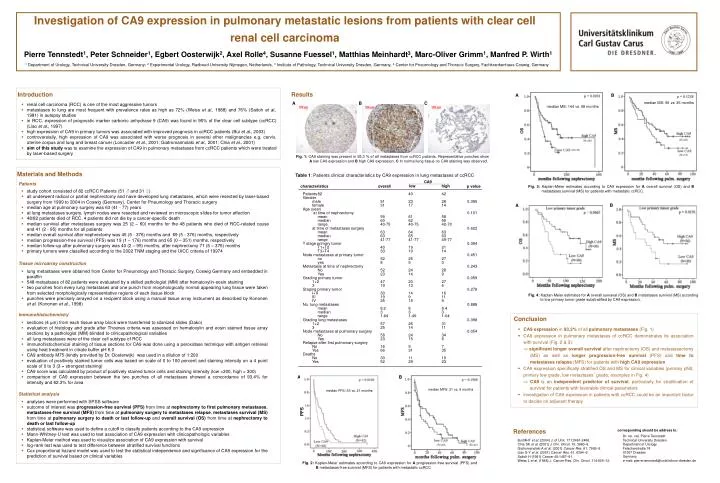
B. A. median MS: 56 vs . 35 months. A. B. C. median MS: 144 vs . 58 months. (N=40). (N=40). (N=37). (N=37).

E N D
B A median MS: 56 vs. 35 months A B C median MS: 144 vs. 58 months (N=40) (N=40) (N=37) (N=37) Fig. 1: CA9 staining was present in 93,3 % of all metastases from ccRCC patients. Representative punches show A low CA9 expression and B high CA9 expression. C In normal lung tissue no CA9 staining was observed. Materials and Methods Patients Table 1: Patients clinical characteristics by CA9 expression in lung metastases of ccRCC • study cohort consisted of 82 ccRCC Patients (51 ♂ and 31 ♀) • all underwent radical or partial nephrectomy and have developed lung metastases, which were resected by laser-based surgery from 1999 to 2004 in Coswig (Germany), Center for Pneumology and Thoracic surgery • median age at pulmonary surgery was 63 (41 - 77) years • at lung metastases surgery, lymph nodes were resected and reviewed on microscopic slides for tumor affection • 48/82 patients died of RCC, 4 patients did not die by a cancer-specific death • median survival after metastases surgery was 25 (2 – 60) months for the 48 patients who died of RCC-related cause and 41 (2 - 95) months for all patients • median overall survival after nephrectomy was 45 (5 - 376) months and 69 (5 - 376) months, respectively • median progression-free survival (PFS) was 19 (1 – 176) months and 65 (0 – 351) months, respectively • median follow-up after pulmonary surgery was 40 (2 – 95) months, after nephrectomy 71 (5 – 376) months • primary tumors were classified according to the 2002 TNM staging and the UICC criteria of 19974 Fig. 3: Kaplan-Meier estimates according to CA9 expression for A overall survival (OS) and B metastases survival (MS) for patients with metastatic ccRCC. CA9 low high characteristics overall p value Patients 82 40 42 Gender male 51 23 28 0.395 female 31 17 14 Age (year) at time of nephrectomy 0.101 mean 59 61 58 median 60 62 59 range 40-75 40-75 42-70 at time of metastases surgery 0.622 mean 63 64 63 median 63 65 63 range 41-77 41-77 49-77 T stage primary tumor 0.394 T1+T2 40 19 21 T3+T4 33 19 14 Node metastases at primary tumor 0.451 no 52 25 27 yes 8 5 3 Metastasis at time of nephrectomy 0.243 No 52 24 28 Yes 23 14 9 Grading primary tumor 0.059 1+2 47 20 27 3 19 13 6 Staging primary tumor 0.278 I+II 30 14 16 III 19 9 11 IV 25 15 9 No. lung metastases 0.888 mean 8.2 8 8.4 median 3 3 3 range 1-64 1-49 1-64 Grading lung metastases 0.398 1+2 57 26 31 3 25 14 11 Node metastases at pulmonary surgery 0.054 No 58 24 34 Yes 23 15 8 Relapse after first pulmonary surgery No 16 9 7 Yes 66 31 35 Deaths No 30 11 19 Yes 52 29 23 B A Tissue microarray construction • lung metastases were obtained from Center for Pneumology and Thoracic Surgery, Coswig Germany and embedded in paraffin • 548 metastases of 82 patients were evaluated by a skilled pathologist (MM)after hematoxylin-eosin staining • two punches from every lung metastases and one punch from morphologically normal appearing lung tissue were taken from selected morphologically representative regions of each tissue block • punches were precisely arrayed on a recipient block using a manual tissue array instrumentas described by Kononen et al. (Kononen et al., 1998) Immunohistochemistry • sections (4 µm) from each tissue array block were transferred to silanized slides (Dako) • evaluation of histology and grade after Thoenes criteria was assessed on hematoxylin and eosin stained tissue array sections by a pathologist (MM) blinded to clinicopathological variables • all lung metastases were of the clear cell subtype of RCC • immunohistochemical staining of tissue sections for CA9 was done using a peroxidase technique with antigen retrieval using heat treatment in citrate buffer pH 6.0 • CA9 antibody M75 (kindly provided by Dr. Oosterwijk)was used in a dilution of 1:200 • evaluation of positively stained tumor cells was based on scale of 0 to 100 percent and staining intensity on a 4 point scale of 0 to 3 (3 = strongest staining) • CA9 score was calculated by product of positively stained tumor cells and staining intensity (low <300, high = 300) • comparison of CA9 expression between the two punches of all metastases showed a concordance of 93.4% for intensity and 62.3% for area A B Statistical analysis • analyses were performed with SPSSsoftware • outcome of interest was progression-free survival(PFS) from time at nephrectomy to first pulmonary metastases, metastases-free survival(MFS) from time at pulmonary surgery to metastases relapse, metastases survival (MS) from time at pulmonary surgery to death or last follow-up and overall survival (OS) from time at nephrectomy to death or last follow-up • statistical software was used to define a cutoff to classify patients according to the CA9 expression • Mann-Whitney-U test was used to test association of CA9 expression with clinicopathologic variables • Kaplan-Meier method was used to visualize association of CA9 expression with survival • log-rank test was used to test difference between stratified survival functions • Cox proportional hazard model was used to test the statistical independence and significance of CA9 expression for the prediction of survival based on clinical variables median MFS: 21 vs. 9 months median PFS: 53 vs. 21 months PFS (N=40) (N=42) Fig. 2: Kaplan-Meier estimates according to CA9 expression for A progression-free survival (PFS) and B metastases-free survival (MFS) for patients with metastatic ccRCC Investigation of CA9 expression in pulmonary metastatic lesions from patients with clear cell renal cell carcinoma Pierre Tennstedt1, Peter Schneider1, Egbert Oosterwijk2, Axel Rolle4, Susanne Fuessel1, Matthias Meinhardt3, Marc-Oliver Grimm1, Manfred P. Wirth1 1 Department of Urology, Technical University Dresden, Germany; 2 Experimental Urology, Radboud University Nijmegen, Netherlands, 3 Institute of Pathology, Technical University Dresden, Germany, 4 Center for Pneumology and Thoracic Surgery, Fachkrankenhaus Coswig, Germany Introduction Results A B • renal cell carcinoma (RCC) is one of the most aggressive tumors • metastases to lung are most frequent with prevalence rates as high as 72% (Weiss et al., 1988)and 76% (Saitoh et al., 1981)in autopsy studies • in RCC, expression of prognostic marker carbonic anhydrase 9 (CA9) was found in 95% of the clear cell subtype (ccRCC) (Liao et al., 1997) • high expression of CA9 in primary tumors was associated with improved prognosis in ccRCC patients (Bui et al., 2003) • controversially, high expression of CA9 was associated with worse prognosis in several other malignancies e.g. cervix, uterine corpus and lung and breast cancer (Loncaster et al., 2001; Giatromannolaki et al., 2001; Chia et al., 2001) • aim of this study was to examine the expression of CA9 in pulmonary metastases from ccRCC patients which were treated by laser-based surgery Fig. 4: Kaplan-Meier estimates for A overall survaval (OS) and B metastases survival (MS) according to low primary tumor grade substratified by CA9 expression. Conclusion • CA9 expression in 93,3% of all pulmonary metastases (Fig. 1) • CA9 expression in pulmonary metastases of ccRCC demonstrates its association with survival (Fig. 2 & 3): significantlonger overall survival after nephrectomy (OS) and metastasectomy (MS) as well as longer progression-free survival (PFS) and time to metastases relapse (MFS) for patients with high CA9 expression • CA9 expression specifically stratified OS and MS for clinical variables (primary pN0, primary low grade, low metastases´ grade; examples in Fig. 4) CA9 is an independent predictor of survival, particularly for stratification of survival for patients with favorable clinical parameters • investigation of CA9 expression in patients with ccRCC could be an important factor to decide on adjuvant therapy References corresponding should be address to: Dr. rer. nat. Pierre Tennstedt Technical University Dresden Department of Urology Fetscherstraße 74 01307 Dresden Germany e-mail: pierre.tennstedt@uniklinikum-dresden.de Bui MHT et al. (2004) J. of Urol. 171:2461-2466. Chia SK et al. (2001) J Clin. Oncol. 19: 3660–8. Giatromanolaki A et al. (2001) Cancer Res. 61: 7992–8. Liao S-Y et al. (2001) Cancer Res. 61: 6394–9. Saitoh H (1981) Cancer 48:1487–91. Weiss L et al. (1988) J. Cancer Res. Clin. Oncol. 114:605–12.