Download

1 / 57
600 likes | 1.11k Vues
Examination of the Heart. Examination of the Heart.

E N D
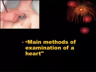
Examination of the Heart
Examination of the Heart In the present era of technological advances, particularly in the various imaging modalities, there is a growing conception among practicing physicians in cardiovascular medicine that bedside physical examination is unnecessary and does not provide useful information.
It should be emphasized, however, that for proper application and interpretation of various new and old tests that are available for cardiovascular evaluation in a given patient.
Bedside clinical examination should be performed and practiced in the same way following similar sequences.
Preparing the patient The heart examination should be made as easy as possible for the patient, who usually expects it to be a relatively distasteful experience. If the physician is considerate and gentle, the patient should feel when it is all over, that most of his or her fears on that score were unfounded.
The ideal examining room is private, warm enough to avoid chilling, and free from distracting noise and sources of interruption. Adequate (preferably fluorescent or natural) light is essential.
The examining table may be placed with its head against the wall, but both sides (particularly the right) and the foot should be accessible to the examiner. And the results should be recorded carefully.
Landmarks and topographic anatomy Certain basic landmarks midsternal line(前正中线) midclavicular lines(锁骨中线) anterior, middle, and posterior axillary lines(腋前、中、后线)
suprasternal notch(胸骨上窝) identification of various ribs and intercostal space precordium(心前区)
Inspection Inspection of the precordium should begin at the foot of the bed. The subject should be supine with the leg horizontal and the head and trunk elevated to approximately 15-30 degrees.
Asymmetry of the thoracic cage due to a convex bulging of the precordium suggests the presence of heart disease since childhood, such as congenital heart disease and rheumatic heart disease, with skeletal molding to accommodate cardiac enlargement.
In the adult, precordial bulge may be produced from the massive pericardial effusion(心包积液).
apical impulse(心尖搏动) Most part of apex is left ventricle. The apex strikes the chest during systole.
The apex impulse is normally located in or about the fifth costal interspace inside the left midclavicular line when the patient is supine. The extent of impulse is about 2~2.5 cm.
Normal apical impulse : It’s location duration intensity amplitude
Usually it is detectable in only one intercostal space and is less than 2-2.5 cm in diameter. The normal apex impulse is characterized by a brief early systolic out ward thrust of moderate amplitude, which ends before the second heart sound.
The apical impulse is normally exaggerated in thin, young individuals and when the subject is in the left lateral decubitus position(左侧卧位).
When a patient takes a deep inspiration and holds his breath, the apical impulse moves downward from the fifth to the sixth interspace.
When the patient lies on his right side, it moves slightly toward the right (1~ 2.5cm), and when he lies on his left side it moves about 2~3 cm toward the left.
The absence of mobility leads one to suspect an adherent pericardium. However, a deep inspiration may bring the lungs over the heart so that the impulse disappears altogether.
Diastolic movements are not perceptible in most cases, but in children and young adults an early diastolic F wave is occasionally present.
Displacement of the apical impulse Heart disease Thoracic disease Abdominal disease
Heart disease Some heart diseases cause the left ventricular dilatation(增大), the apical impulse is displaced laterally and inferiorly and sustained ,
and it may be shifted to the left and upward in right ventricular dilatation .
In aortic disease the impulse is displaced both laterally and downward.
It can be found at the right fifth intercostal space in dextrocardia(右位心) and can not be found in massive pericardial effusion.
Thoracic disease Pneumothorax(气胸) and pleural effusion(胸腔积液) will displace the apical impulse to the normal side. Pleural-adhesion(胸膜粘连) and atelectasis(肺不张) will result in a displacement of impulse toward the diseased side.
Effect of massive right pleural effusion or pneumothorax
The examiner should always observe the shape and contour of patient’s chest. Depressions of the sternum, Kyphosis of dorsal spine(驼背), scoliosis(脊柱侧凸) often alter the shape and position of the apical impulse.
Abdominal disease The apical impulse also can be displaced by large mass(肿瘤), massive ascites(腹水).
The apical impulse may have increased amplitude and duration in those persons with a thin chest, anemia(贫血), fever, hyperthyroidism (甲亢) and anxiety.
Inward impulse(负性心尖搏动): the apex depress far from the chest instead of strikes the chest during systole. Broadbent’s sign is of value in the diagnosis of adherent pericardium(粘连性心包炎). It is also seen in RVH.
Abnormal pulsations in the other areas: Right vertricular hypertophy (RVH). The impulse is clearly seen in left third fourth intercostal space.
Pulmonary emphysema(肺气肿) with RVH, usually the pulsation can be found inferior the xiphoid process(剑突下搏动).
In ascending or arch aortic aneurysm(主动脉瘤), one may detect abnormal pulsations in aortic area, with bulging or pulsation in systole.
Pulmonary hypertension with dilatation the pulsation in systole may be detected in left second intercostal space to the edge of sternum.
Marked pulsation at the base of the heart is seen in aortic insufficiency(主闭), in a dilated aorta or a saccular aneurysm.
Precordial bulge (心前区隆起) congenital heart disease rheumatic heart disease (before puberty) pericardical effusion (adult life) Review
Normal apical impulse The apex impulse is normally located in or about the fifth costal interspace inside the left midclavicular line when the patient is supine. The extent of impulse is about 2~2.5 cm.
Displacement of the apical impulse Heart disease LVD displaced to lateral and inferior
Displacement of the apical impulse RVD displaced to left and upward
Displacement of the apical impulse Congenital dextrocardiac right CHF, myocarditis, myocardiopathy apical impulse decrease intensity
Displacement of the apical impulse Massive pericardial effusion apical impulse disappear
Displacement of the apical impulse Thoracic disease pneumothorax, pleural effusion shifted to healthy side