Download
1 / 61
610 likes | 951 Vues
3 th EU-Meeting on Cobalamins and Mimics Antwerp - Belgium. Introduction to Mass Spectrometry. Eddy Esmans May 2004. I. Introduction. II. Ionization methods. 1. Electron impact. 2. Chemical ionization and DCI, NICI. 3. FAB, SIMS, LD and MALDI. 4. Field desorption.

E N D
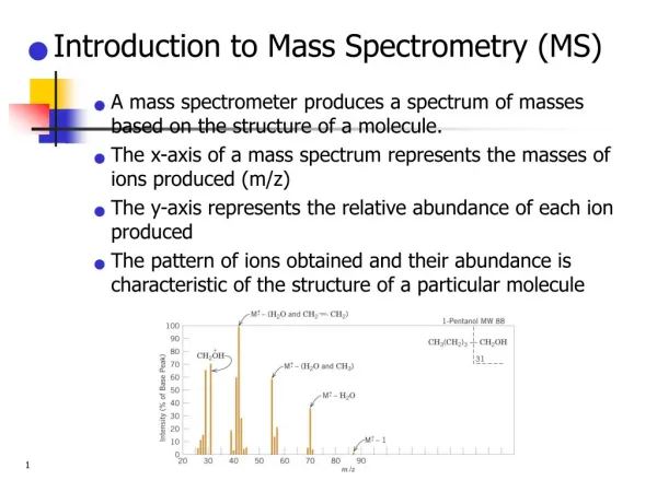
3th EU-Meeting on Cobalamins and Mimics Antwerp - Belgium Introduction to Mass Spectrometry Eddy Esmans May 2004
I. Introduction II. Ionization methods 1. Electron impact 2. Chemical ionization and DCI, NICI 3. FAB, SIMS, LD and MALDI 4. Field desorption 5. Electrospray ionization III. Analyzers 1. Magnetic sector 2. Quadrupole – ion trap 3. Fourier transform 4. Time of flight IV. MS/MS-methods
Ion Detector Ion Source Analyzer Inlet system The Components of a Mass Spectrometer m/z Mass Spectrum Computer
I. Introduction II. Ionization methods 1. Electron impact 2. Chemical ionization and DCI, NIC 3. FAB, SIMS, LD and MALDI 4. Field desorption 5. Electrospray ionization III. Analysers 1. Magnetic sector 2. Quadrupole – ion trap 3. Fourier transform 4. Time of flight IV. MS/MS-methods
E – E0 E II. Ionization methods 1. Electron impact kr E M+. Fi+. 70 eV electron Unimolecular type E = internal energy of e.g. M+. N-1 E0 = activation energy of a particular fragmentation kr = n N = degrees of freedom n = frequency factor IONIZATION EFFICIENCY : ca. 1/1000
A polyatomic molecule does not fragment immediately but during ionization period of 10-16 sec it undergoes a few vibrations. • fragmentation is a “relative slow” process. QET : Quasi Equilibrium Theory b. The energy transferred to M is not localised but is statistically spread over the molecule. c. If the event occurs than this energy is concentrated at one particular bond. This bond will break here. d. The probability of breaking a particular bond in not a function of abundance. e. Metastable ions are formed : ions with a life time of > 10-6 seconds.
Interconversion n”5 n”4 n”3 n”2 n’5 n”1 n’4 n”0 n’3 n’2 AB+. n’1 n’0 AB+. -Ip (theoretical) n3 n3 n3 n2 n1 n0 AB E-impact-ionisation occurs according to the Frank-Condon-principle (vibration is 100 times slower than ionisation)
I. Introduction II. Ionization methods 1. Electron impact 2. Chemical ionization and DCI, NICI 3. FAB, SIMS, LD and MALDI 4. Field desorption 5. Electrospray ionization III. Analysers 1. Magnetic sector 2. Quadrupole – ion trap 3. Fourier transform 4. Time of flight IV. MS/MS-methods
2. Chemical ionization (CI) and DCI, NICI M(g) + reagent gas [MH]+ benefit: producing molecular mass information proton affinity !!! proton affinity PA of M > proton affinity PA of the reacting species Classical reagent gasses: Methane: CH5+ NH3: NH4+ (NH3+. + NH3 NH4+ + NH2.) Isobutane: C4H9+ PS : if PA(M) PA(reagent gas) [MH]+ + ADDUCT FORMATION [M + NH4]+ [M + C2H5]+ if PA(M) < PA(reagent gas) only adducts bad sensitivity
Negative Ion Chemical Ionization Principle : ion souce is filled with CH4 and 70 eV electrons are slowed down to thermal energy. These electrons can be “captured” by molecules containing sulphur (cfr. Electron capture GC) formation of M°--ions
I. Introduction II. Ionization methods 1. Electron impact 2. Chemical ionization and DCI, NICI 3. FAB, SIMS, LD and MALDI 4. Field desorption 5. Electrospray ionization III. Analysers 1. Magnetic sector 2. Quadrupole – ion trap 3. Fourier transform 4. Time of flight IV. MS/MS-methods
Fast atom bombardment (FAB) and Secundary Ion Mass Spectrometry (SIMS) Ions (Cs+) Neutrals (Ar, Xe, …) Principle: IONS analysed Sample 1. FAB : Ar + e Ar+ acceleration (5-15 KeV) Ar+ + Ar Ar + Ar+ fast slow + 8 KeV slow fast 2. SIMS : Cs+ generated (35 KeV) 3. LSIMS : Sputtering yield (number of particles ejected/incident particle) Dependent on mass and velocity of impinging particle
Matrix properties 1. Good solubility 2. Vapour pressure must be sufficiently low to maintain vacuum conditions 3. Viscosity must allow diffusion of the analyte from the bulk to the surface 4. Polar : to solvate and separate preformed ion glycerol, 3-nitrobenzylalcohol, mixture of 1,4-dithiothreitol/1,4-dithioerythitol 5:1 (magic bullet)
Laser Desorption & Matrix Assisted Laser Desorption A few lasers: N2 –laser : 337 nm Nd-Yag laser : 354 & 266 nm E: 20mJ/cm2
I. Introduction II. Ionization methods 1. Electron impact 2. Chemical ionization and DCI, NICI 3. FAB, SIMS, LD and MALDI 4. Field desorption 5. Electrospray ionization III. Analysers 1. Magnetic sector 2. Quadrupole – ion trap 3. Fourier transform 4. Time of flight IV. MS/MS-methods
I. Introduction II. Ionization methods 1. Electron impact 2. Chemical ionization and DCI, NICI 3. FAB, SIMS, LD and MALDI 4. Field desorption 5. Electrospray ionization III. Analysers 1. Magnetic sector 2. Quadrupole – ion trap 3. Fourier transform 4. Time of flight IV. MS/MS-methods
Picofrit columns™ • injection: 1 ml • flow-rate: 500 nl/min • isocratic 20/80 NH4Ac (0.01 M) / MeOH • column: AQUASIL C18, 75 mm x 4.9 cm (15cm 2cm), tip 5 mm
I. Introduction II. Ionization methods 1. Electron impact 2. Chemical ionization and DCI, NICI 3. FAB, SIMS, LD and MALDI 4. Field desorption 5. Electrospray ionization III. Analyzers 1. Magnetic sector 2. Quadrupole – ion trap 3. Fourier transform 4. Time of flight IV. MS/MS-methods
I. Ion source : Ions get kinetic energy V 8 KV V = tension m = mass v = speed z = charge
II. Electrostatic sector : E = electrostatic field = Ions with the same Ekin will travel with the same r and leave the electrostatic sector at the same point (This is independant of their mass !!!)
I. Introduction II. Ionization methods 1. Electron impact 2. Chemical ionization and DCI, NIC 3. FAB, SIMS, LD and MALDI 4. Field desorption 5. Electrospray ionization III. Analyzers 1. Magnetic sector 2. Quadrupole – ion trap 3. Fourier transform 4. Time of flight IV. MS/MS-methods
a. Quadrupole filter E = E0 (x + y + z) Quadrupole field: Independent field in x,y,z-directions. Ionsentering this field will undergo a force F = eE
Quadrupole field subjected to the restraites imposed by the Laplace-equations: Physical meaning : the Laplacean is a measure for the distorsion of the E-field
Applied potential 1. Equation of motion of the ions entering this field mx = eEx ¨ - mx ¨ mx ¨ ¨ x xz and yz motion of ions in plane ¨ y mz = 0 ¨ Velocity in z-direction is cte but ions are accelerated in x and y-directions !
Stability diagram 0 = U + V.cost ( = 2f) ¨ x ¨ y Matthieu-equations U = 500-2000 V V = 0-3000 V
Stability diagram scanning : changing U and V but keeping what if U = 0 resolution = 0 Rf-quadrupole only will be able to pass m/z-values > certain m/z-value as long as V is in stability area.
I. Introduction II. Ionization methods 1. Electron impact 2. Chemical ionization and DCI, NIC 3. FAB, SIMS, LD and MALDI 4. Field desorption 5. Electrospray ionization III. Analyzers 1. Magnetic sector 2. Quadrupole – ion trap 3. Fourier transform 4. Time of flight IV. MS/MS-methods
Ion cyclotron resonance Fourier Transform MS Ion can be trapped in a H-field The ion will have a stable trajectory when : circular motion with frequency Relation between and m/z-value each m/z-value will move with its typical frequency/radius
Simultaneously excite all ions by electromagnetic pulse (1µs). Depending on their m/z-values ions will absorb energy at their frequency and subsequently get hifgher trajectories close to the receive plates. All the frequencies detected in this time ellaps by the receive plates at the same time.
I. Introduction II. Ionization methods 1. Electron impact 2. Chemical ionization and DCI, NIC 3. FAB, SIMS, LD and MALDI 4. Field desorption 5. Electrospray ionization III. Analyzers 1. Magnetic sector 2. Quadrupole – ion trap 3. Fourier transform 4. Time of flight IV. MS/MS-methods
Resolution > 10.000 Mass range 50.000 Time of Flight Source : ion flight tube : L
Time of Flight Reflectron : corrects for energy dispension
Time an ion spends in reflectron correct energy E 2 ions with mass M energy E’ t, t’ = flight time in the field free region of TOF
Ions come in the reflectron : penetrate a distance x or x’ x’ = a2x Conclusion : a>1 E’kin>Ekin t’flight<tflight but x’ > x a<1 E’kin< Ekin t’flight>tflight but x’ < x