Download
1 / 75
760 likes | 1.09k Vues
The Cardiovascular System. The major organs of the cardiovascular system The heart structure and function. After today you should be able to: For more help: Chapter13 pp. 329-364. Name the organs of the cardiovascular system and discuss their functions.

E N D
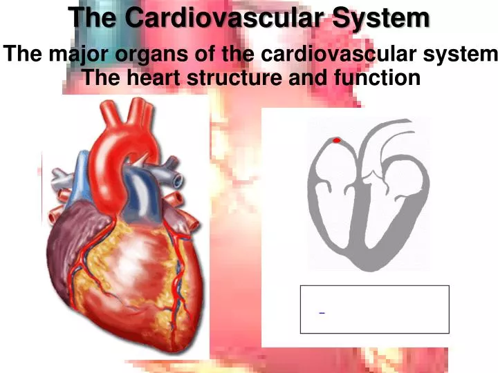
The Cardiovascular System The major organs of the cardiovascular systemThe heart structure and function
After today you should be able to: For more help: Chapter13 pp. 329-364 • Name the organs of the cardiovascular system and discuss their functions. 2. Name and describe the locations and functions of the major parts of the heart. 3. Trace the pathway of the blood through the heart and the vessels of the coronary circulation.
Major organs of the cardiovascular system • The heart– located in the pericardial cavity, slightly to the left, close to the left lung, and rests on the diaphragm • Arteries – strong elastic vessels that are adapted for carrying blood away from the heart under high pressure
Major organs of the cardiovascular system • Arterioles– smaller branches coming from the arteries • Capillaries- smallest of the artery system, connect the smallest arterioles and the smallest venules
Major organs of the cardiovascular system • Venules– the smallest vessels of the venous system, that continue from the capillaries and merge to form veins • Veins- carry blood back to the atria of the heart following pathways that are almost parallel to the arteries. Similar to arteries, but have thinner walls, and generally have flap like valves. Generally lower pressure than that of the arteries.
The Heart • In the course of a lifetime, a human heart can beat over two million times. • It can carry 7,000 liters of blood throughout the body. • Composed of cardiac muscle tissue • The wall of the heart or tissue is made up of three levels: outer most: epicardium, middle: myocardium and inner: endocardium
The Heart • The heart is divided into four chambers: • The LEFT and RIGHT ATRIA • The LEFT and RIGHT VENTRICLES • A sophisticated valve system controls blood flow between the chambers. In fact, it is the latching (when they open and close) of the heart valves that creates the beating sound of the heart. • There are four distinct valves.
The Heart • The four chambers:
The Heart • The valves:
Superior Vena Cava The superior vena cava is one of the two main veins bringing de-oxygenated blood from the body to the heart. Veins from the head and upper body feed into the superior vena cava, which empties into the right atrium of the heart. Why is the blood here de-oxygenated?
Inferior Vena Cava • Inferior vena cava :is one of the two main veins bringing de-oxygenated blood from the body to the heart. • Veins from the legs and lower torso feed into the inferior vena cava, which empties into the right atrium of the heart.
Right Atrium • The right atrium receives de-oxygenated blood from the body through the superior vena cava and inferior vena cava .
Tricuspid Valve • Tricuspid valve:separates the right atrium from the right ventricle. • It opens to allow the de-oxygenated blood from the right atrium to flow into the right ventricle. • It closes as the right ventricle contracts, preventing blood from returning to the right atrium. • Forces blood to exit into the pulmonary artery.
Right Ventricle • The right ventricle: receives de-oxygenated blood as the right atrium contracts. • This blood will move from the right ventricle to the pulmonary artery leading to the lungs.
Pulmonary Valve • Pulmonary valve: separates the right ventricle from the pulmonary artery. • As the ventricles contract, it opens to allow the de-oxygenated blood from the right ventricle to flow to the lungs. • It closes as the ventricles relax, preventing blood from returning to the heart.
Pulmonary Artery • Pulmonary artery: is the vessel transporting de-oxygenated blood from the right ventricle to the lungs. • A common misconception is that all arteries carry oxygen-rich blood. • It is more appropriate to classify arteries as vessels carrying blood away from the heart.
Summarize what we know so far: ANSWER THE FOLLOWING QUESTIONS: • Where does blood originate from? • What in the heart does it enter first? • Where does it go next? • What are the roles of the 2 valves? • Where does blood exit and go to from the right side of the heart? • Is it de-oxygenated (oxygen poor) or oxygenated (oxygen rich)?
The Heart: Left side • Brings oxygenated (oxygen rich blood) from the lungs to the heart. • Begins with the Pulmonary Vein.
Pulmonary Vein • Pulmonary vein: is the vessel transporting oxygen-rich bloodfrom the lungs to the left atrium. • A common misconception is that all veins carry de-oxygenated blood. • It is more appropriate to classify veins as vessels carrying blood to the heart.
Left Atrium • Left atrium: receives oxygenated blood from the lungs through the pulmonary vein. • As the heart contracts (triggered by the sinoatrial node) Blood travels through the atria. • It passes through the mitral valve into the left ventricle.
Mitral (Bicuspid) Value • Mitral valve: separates the left atrium from the left ventricle. • It opens to allow the oxygenated blood in the left atrium to flow into the left ventricle. • It closes as the left ventricle contracts, preventing blood from returning to the left atrium. • Forcing it to exit through the aortic valve into the aorta.
Left Ventricle • Left ventricle: receives oxygenated blood as the left atrium contracts. • The walls of the left ventricle are thicker than the walls of the right ventricle, so that they can generate enough force to push the blood from the left ventricle into the aorta.
Aortic Valve • Aortic valve: separates the left ventricle from the aorta. • As the ventricles contract, it opens to allow the oxygenated blood collected in the left ventricle to flow throughout the body. • It closes as the ventricles relax, preventing blood from returning to the heart.
Aorta • Aorta: is the largest single blood vessel in the body. • It is approximately the diameter of your thumb. • This vessel carries oxygen-rich blood from the left ventricle to the various parts of the body.
Papillary Muscles • Papillary muscles: attach to the lower portion of the interior wall of the ventricles. • They connect to the chordae tendineaeon the valves, • The contraction of the papillary muscles opens the valves. When the papillary muscles relax, the valves close.
Chordae Tendineae • Chordae tendineaeare tendons linking the papillary muscles to the tricuspid valve in the right ventricle and the mitral valve in the left ventricle. • The chordae tendineae are string-like in appearance and are sometimes referred to as "heart strings."
Ventricular Septum • Ventricular Septum: wall separating the lower chambers (the ventricles) of the heart from one another.
As a group of 4: • With the construction paper: Create the heart. • Show the major organs that we discussed (arteries, veins, valves, chambers, etc) • Show the pathway that blood takes • Make sure the red and blue construction paper are in the correct location for oxygenated and de-oxygenated. • Use the white and black construction paper to show where CO2 is and goes and where O2 is and goes. • The shape needs to look similar to the actual heart.
Act out the blood flow through the circulatory and respiratory system card activity
serves as the natural pacemaker for the heart. Nestled in the upper area of the right atrium, it sends the electrical impulse that triggers each heartbeat. The impulse spreads through the atria, coordinated wave-like manner. Sinoatrial Node (often called the SA node or sinus node)
The impulse that originates from the SA node strikes AV node situated in the lower portion of the right atrium. The AV node in turn sends an impulse through the nerve network to the ventricles to contract. Atrioventricular node (or AV node)
electrical network serving the upper ventricles These nerve fibers send impulses that cause the cardiac muscle tissue to contract. Right and Left Bundle Branches.
electrical network serving the lower ventricles These nerve fibers send impulses that cause the cardiac muscle tissue to contract. Purkinje Fibers
Electrical Conduction Pathway: • The SA Node to the AV Node to the left and right Bundle Branches - to the Purkinje Fibers = THE HEART BEAT and CONTRACTIONS
BLOOD • Blood is a mixture of Cells and Plasma • The heart pumps blood through arteries • Blood carries oxygen to the body and wastes away from the body.
Blood Cells: Contains 3 types of Cells: • RED BLOOD CELLS • WHITE BLOOD CELLS • PLATELETS
Blood Cells: Identify the components: white blood cell plasma platelets
Red Blood Cells: Erythrocytes • Biconcave discs that allows it to transport gases • Hemoglobin binded to oxygen gives it the red color. • When they are mature they lack nuclei. • RBC count for adults is: 4,600,000-6,200,000 cells per mm3 • 120 day life span • Made in the bone marrow
White Blood Cells: Leukocytes • Protect against disease • Part of the Immune system • Twice the size of red blood cells. • WBC count: 4,000-10,000 normally • During an infection this number increases rapidly. After the infection goes back to normal.
White Blood Cells: Leukocytes • Six different types: * Neutrophils – 58% *Eosinophils- 2 % * Basophils – 1% *Bands - 3 % • * Monocytes - 4 % * Lymphocytes - 4 %
Platelets: Thrombocytes • Only fragments of cells that have broken off from Megakaryocytes in the bone marrow. • Their main function is in blood clotting. • Because of their function they contain: chemicals such as epinephrine, cytokines, and others that aid in blood clotting. • Ten day life span • VERY SMALL! • Platelet cell count: 130,000-360,000