Download

1 / 22
240 likes | 704 Vues
The typical spinal nerve. Medical Biomorphology 140 Stuart Bunt smbunt@anhb.uwa.edu. . Order of the lecture. Embryology Evolution Anatomy Dermatomes Peripheral nerve territories Autonomics. Somites (blocks of mesoderm). Somites appear early in development

E N D
The typical spinal nerve Medical Biomorphology 140 Stuart Bunt smbunt@anhb.uwa.edu.
Order of the lecture • Embryology • Evolution • Anatomy • Dermatomes • Peripheral nerve territories • Autonomics
Somites (blocks of mesoderm) • Somites appear early in development • Are characteristic of all vertebrates
Somites are found in all vertebrates Chicken Embryos
In humans the segments are most obvious in the vertebrae and segmental nerves of the thoracic area.
Somitic tissue migrates to form muscles The somite blocks disperse and the mesodermal cells contribute to the dermis, muscles, bones and connective tissue.
Somite splits in to dermo-myotome and the dispersing cells of the sclerotome • The sclerotome gives rise to the connective tissue and tissue derived from it such as the cardiovascular system and skeletal system • The dermo-myotome splits in to dermal and myotomal components • The former forms the deep layers of the dermis • The myotome forms the muscles
The myotome forms a dorsal epimere and a ventral hypomere Epimere forms vertebral musculature, hypomere the rest of the muscles of the body wall and limbs
While the somites are forming the neural tube closes The crest of the neural tube migrates off in to the body These cells form the dorsal root ganglia and contribute to many other tissues from the facial skeleton to the adrenals
Nerves grow out in to the mesoderm Nerve fibres follow surface clues (glyco proteins), entering the front of each myotome As limb buds grow the fibres extend and innervate overlapping but regular areas of the limb.
The area innervated by fibres from a single root is known as a DERMATOME
The area innervated by a single peripheral nerve is NOT the same as a Dermatome. If you look carefully at the dermatome patterns in different books you will see that they often disagree. This is because the patterns are variable and overlap considerably
Further growth gives rise to the adult pattern of a typical spinal nerve Dorsal ramus Ventral ramus
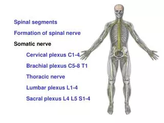
Spinal nerves • 8 Cervical • 12 thoracic • 5 lumbar • 5 sacral • 1 coccygeal Each segment made of tissue from one somite
Dorsal root • Contains sensory fibres • Cell bodies in the dorsal root ganglion • Runs in to dorsal horn of spinal cord • Distal process carries sensation from the periphery
Dorsal Root Ganglion • contains pseudounipolar sensory cells • Schwann cells • Satellite cells • NO synapses
Ventral Root • somatic motor axons • visceral motor axons • Cell bodies in spinal cord grey matter • terminals on muscles or in autonomic ganglia
The spinal nerve • Only part that contains ALL segmental nerves • Motor AND sensory • Short segment just outside vertebral canal
Dorsal and Ventral Primary Rami • Dorsal Ramus supplies • superficial structures on dorsal midline of body • skin, skeleton, muscles, meninges on post vert column • Ventral Ramus supplies • deep and superficial structures of lateral and anterior trunk • skin, skeleton, muscles, meninges
Grey and White Rami Communicantes • The grey rami contain unmyelinated visceral axons • heading out from the ganglia to the periphery • postganglionic axons • The white rami contain myelinated fibres (white) • Coming from the spinal cord to the ganglia • preganglionic axons • T1-L2
The sympathetic ganglia • Visceral autonomic ganglia • Postganglionic fibres innervate gut etc. • Connected by connectives • Run alongside the vertebral column
Splanchnic Nerves • Innervate internal organs • preganglionic and postganglionic • sympathetic or parasympathetic