Download
1 / 61
710 likes | 1.71k Vues
Dag 3 Ischemie en infarct. Martijn Meuwissen Academisch Medisch Centrum, Amsterdam. non-profit / open access / physician moderated / up-to-date. Cursusoverzicht. Dag 1: Basis, systematische beoordeling Ivo van der Bilt Dag 2: Ritmestoornissen Jonas de Jong Dag 3: Ischemie

E N D
Dag 3Ischemie en infarct Martijn Meuwissen Academisch Medisch Centrum, Amsterdam
Cursusoverzicht • Dag 1: Basis, systematische beoordeling • Ivo van der Bilt • Dag 2: Ritmestoornissen • Jonas de Jong • Dag 3: Ischemie • Martijn Meuwissen De cursus is interactief. Onderbreek gerust!
Cardionetworks Auteurs: • Jonas de Jong • Ivo van der Bilt • Martijn Meuwissen • Renée van den Brink • Joris de Groot Illustraties: • Rob Kreuger • Bart Duineveld Met dank aan: • Arthur Wilde • Rudolph Koster Boeken: • Wellens: The ECG in Emergency Decision Making • Garcia / Miller: Arrhythmia Recognition • Braunwald Heart Disease
Agenda Ischemie en Infarct • Achtergrond • Diagnostiek • Infarct localisatie • Complicaties • Quiz
Pathofysiologie • Oorzaak acuut coronair syndroom = acute afname of blokkade in coronaire doorbloeding • Atherosclerotische plaque • Thrombotische occlusie tgv plaque ruptuur • Plaatjes aggregatie • Vasoactiva > spasme • Thrombine > stollingsactivatie > bevorderen aggregatie • Zelden: erosie/ embolie/ spasme/ anemie/ hypoxie
Vulnerable Plaque Ruptured Plaque Stable Plaque lumen disrupted fibrous cap thin fibrous cap lumen thick fibrous cap Pathofysiologie lumen adventitia intima thrombus media lipid core macrophages
Levensloop coronaire plaque adhesie en migratie van ontstekingscellen migratie van monocyten en gladde spier cellen schuimcel vorming atheroom en fibreuze cap vorming “vulnerable plaque” – fragiele lesie gecompliceerde lesie 0% 20% 30% 50% 70% 90% tijd
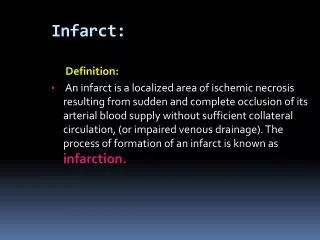
Pathofysiologie • Acuut myocard infarct • Enzymen (troponines, CK-MB) + 1 of meer van de volgende • Ischaemische symptomen • ECG afwijkingen passend bij ischaemie of necrose: ontwikkeling van Q-golven, ST • Pathologische kenmerken van AMI (echo)
Grootte van infarct Demand vs supply: • Supply: • plaats afsluiting (proximaal/ distaal) • duur afsluiting irreversibel na 20-30 min • collaterale doorbloeding • (Hb, PO2) • Demand • zuurstof behoefte myocard • frequentie, RR, contractiliteit
35-day mortality reduction vs treatment delay Waarom snelle infarctdiagnostiek? 80 Absolute benefit per 1000 treated patients 60 40 20 0 Treatment delay (h) BoersmaLancet. 1996
Patency of infarct-related comparing PTCA and Thrombolysis SP King III, Interventional Cardiology 2007 0
Effect of door-to-door time on mortality 1.8 Relative risk of death Relative Risk of In-Hospital Death 1.6 1.4 1.2 1.0 McNamara et al. JACC 2006;47:2180-2186
AHC / AHA Guidelines Primary PCI is “preferred treatment” for ST-elevation acute myocardial infarction if: • Symptoms < 12 hrs • Time from “first medical contact” to start reperfusion therapy 90 min. • Experienced team, high volume center
ESC Guidelines STEMI 2003 STEMI <12 hours after onset of chest pain patient presenting in a hospital with PCI patient presenting in a hospital without PCI ≥ 3 – 12 hours < 3 hours * immediate transfer Thrombolysis failed successful PCI within up to 24 hours available PCI within up to 24 hours not available predischarge ischaemia primary PCI rescue PCI post thrombolysis PCI ischaemia-driven PCI
Decline in in-hospital mortality S. Rasoul, Thesis 2007
Algemene Infarct Diagnostiek • Anamnese • Drukkende retrosternale POB, dyspnoe, radiatie, angst, vegetatief, (geen) reactie op NTG, collaps , risicofactoren • Lichamelijk onderzoek • Vegetatieve verschijnselen, pols, bloeddruk, decompensatio cordis, shock • ECG • ST deviatie, Q’s, ritme-/ geleidingsstoornissen (LBTB) • Lab • troponine, CKMB etc
AlgemeneDiagnostiek • Mogelijkegevolgen van AMI • Dyspneu: backward failure (MI?) • Shock: forward failure (ruptuur?) • Duizelig/collaps: ritme-/ geleidingsstoornissen (VF familieanamnese!) • CVA: LV thrombus/ AFib • NB: frequent geen (duidelijke) klachtenouderen, diabetici!!
Differentiaal diagnostiek • Cardiaal • (pericarditis, mechanische complicatie (stil) AMI, takotsubo) • Vasculair • Type A (B) dissectie, aneurysma • Pulmonaal • longembolie • Gastro-oesophageaal • spasme • Bewegingsapparaat • Tietze
ECG Diagnostiek • Acute coronair occlusie • ST elevatie, (reciproke) depressie • Q-vorming • T-top inversie • Geleiding: Nieuw linker- / rechter bundeltak blok • As-draai • QT verlenging • Ritmestoornissen (VF/ AIVR/ AF/ totaal AVB) • Instabiele angina pectoris • Persisterende of voorbijgaande ST depressie • T golf veranderingen (vlak / inversie / pseudo-normalisatie) • Niet-specifiek / normaal ECG
ECG Diagnostiek • Precordiaal
ECG Diagnostiek • Precordiaal
ECG Diagnostiek • Extremiteiten
Systematische beoordeling • Algemene kenmerken • Ritme • Frequentie • Geleidingstijden • Hartas • P top morfologie • QRS morfologie • ST morfologie • Vergelijking met oud ECG • Conclusie
7+2 STAPPENPLANStap 6: QRS morfologie • Pathologische Q top? • Breedte ≥ 0.04 sec • Diepte > ⅓ van de R • Differentiaal diagnose? • Oud infarct • Cardiomyopathie (HCM, DCM) • COPD • Intraventriculaire geleidingsstoornissen
7+2 STAPPENPLANStap 6: QRS morfologie • R-top progressie? • Overgangs complex in V3, V4 • Normaal zit het overgangs complex (waar de R-top groter wordt dan de S) bij V3 tot V4
7+2 STAPPENPLANStap 6: QRS morfologie • R-top progressie? • Differentiaal diagnose onvoldoende r-top progressie? • RV hypertrofie • COPD, asthma • Voorwand infarct of anteroseptaal infarct • Geleidingsstoornissen (LBBB, Left anticus hemiblok, intraventriculaire geleidings vertraging) • Cardiomyopathie • Thorax afwijking • Normale variant • Precordiale afleidingen verkeerd geplaatst ANAMNESE EN LO/ ZIJN EXTREEM BELANGRIJK VOOR JUISTE INTERPRETATIE VAN HET ECG
Vorm ST Segment concaaf of convex? B A
7+2 STAPPENPLANStap 7: ST-segment • Wanneer spreekt men van een pathologische ST elevatie? • ST elevatie die op het J punt ≥ 1 mm is in afleiding I, II, III, aVL, aVF, V4-V6 • ST elevatie die op het J punt ≥ 2 mm in V1-V3 • ST elevatie treedt op bij: • Transmurale ischemie of transmuraal infarct (STEMI) • Differentiaal diagnose • Pericarditis, LVH, brugada, takotsubo, K+, digitalis,bundeltakblok, CVA, hyperventilatie etc.
Vroege Repolarisatie • Zeer frequente bevinding • “Smiley”configuratie • Overigens gezonde asymptomatische jonge volwassene • Vaak in voorwands afl. • Notching J punt • Geen Q • Geen reciproke ST depressie
7+2 STAPPENPLANStap 7+1: Vergelijk met oud ECG • Verandering ritme? • Nieuw boezemfibrilleren? • Verandering frequentie? • Bradycardie (SB, AV blokmedicatie effect?) of tachycardie ([S]VT) • Verandering geleidingstijden? • PQ tijd (medicatie?); QRS (ischemie, medicatie?) QT tijd (medicatie?) • Verandering hartas? • Verandering geleiding, infarct doorgemaakt? • Nieuwe pathologische Q’s • Verandering geleiding, infarct doorgemaakt, plaatsing elektroden? • Verandering R top progressie precordiaal? • Afname R (infarct, tamponade, plaatsing elektroden?) • Toename R top (LVH, RVH, verandering geleiding intraventriculair) • Verandering ST segment? • Verandering T-top?
Infarct localisatie • Hoofdstamocclusie: • diffuse ST depressie met ST elevatie in AVR. • Zeer hoog risico • Voorwand: • V1-V4. Stroomgebied: LAD. (vaak tachycardy.) • Onderwand: • II, III, AVF. • Stroomgebied: 80% RCA (elevatie III>II; depressie >I dan in AVL), anders RCX (in 20%). (vaak bradycardy) • Rechter ventrikelfinfarct: • ST↑ in V4R. • vullen indien hypotensief • Posterior: • hoge R en ST-depressie in V1-V3 • Lateraal: • elevatie inI, AVL, V6. Stroomgebied: LAD (D-tak)
OnderwandinfarctRCA of RCx? • RCA occlusie • ST elevatie III > II • ST depressie in aVL > I • V4R isoelectrisch of geëleveerd • RV infarct mogelijk • S:R in aVL > 3 • RCX occlusie: • ST elevatie II > III • V4R negatieve T • S:R in aVL < 3
R ZONE OF ISCHEMIA ISCHEMIA CAUSES INVERSION OF T WAVE DUE TO ALTERED REPOLARIZATION P ZONE OF INJURY Q S T ZONE OF INFARCTION R MUSCLE INJURY CAUSES ELEVATION OF S-T SEGMENT P Q T DEATH (INFARCTION) OF MUSCLE CAUSES Q OR QS WAVES DUE TO ABSENCE OF DEPOLARIZATION CURRENT FROM DEAD TISSUE AND OPPOSING CURRENTS FROM OTHER PARTS OF HEART R P T Q DURING RECOVERY (SUBACUTE AND CHRONIC STAGES) S-T SEGMENT OFTEN IS FIRST TO RETURN TO NORMAL, THEN T WAVE, DUE TO DISAPPEARANCE OF ZONES OF INJURY AND ISCHEMIA R T P RECIPROCAL EFFECTS ON OPPOSITVE SIDE OF INFARCT
ECG patterns and LAD ischemia V3 V5 V2 V4 V1 AVL AVR I AVL AVR V6 III II AVF AVF Proximal LAD occlusion. Global ischemia of the whole anterior and septal aspect of the left ventricle. The ST segment vector points in a superior direction, because the anterobasal segment is the dominant ischemic area. Related ECG changes. The superiorly oriented ST vector leads to ST changes, such as ST elevation in lead AVR and V1 with reciprocal ST depression in the inferior leads and in leads V5 and V6. Adaptedfrom Wellens et al.
ECG patterns and LAD ischemia V3 V1 V4 V5 V2 AVL AVR I AVR V6 III II AVF AVF Related ECG changes. The inferiorly directed ST vector leads to ST depression in lead AVR, and ST segment elevation in the inferior leads. Distal LAD occlusion. The ST vector points more inferiorly due to ischemic dominance of the inferoapical area. Adaptedfrom Wellens et al.
ECG patterns and LAD ischemia AVL AVR I III II AVF Perfusion areas of the left anterior descending branch of the left coronary artery (LAD) Myocardial areas perfused by branches from the LAD include the septal, lateral and inferoapical area. The ST segment elevation vector is the result of the amount of ischemia in these respective areas. Possible occlusion sites related to the main branches of the LAD Upper left. Proximal to first septal and first diagonal branch. Lower left. Distal to first septal, proximal to first diagonal branch. Upper right. Distal to first diagonal, proximal to first septal branch. Lower right. Distal to both branches. Adaptedfrom Wellens et al.