Download

1 / 19
210 likes | 435 Vues
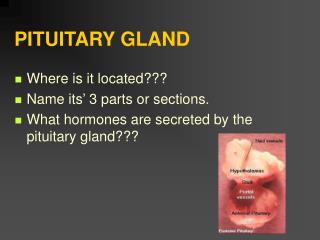
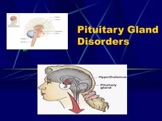
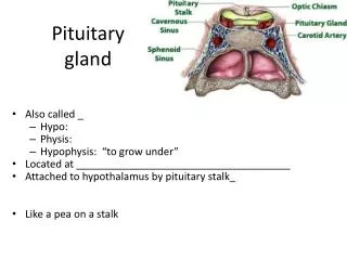
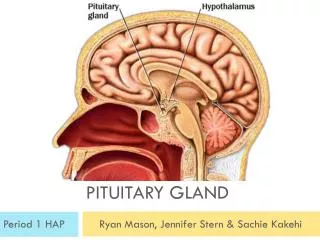
PITUITARY GLAND. Location. A small gland which lies in the hypophyseal fossa hanging from the hypothalamus, to which it is connected. Its dimensions are 1 x 1x 1.5 cm and its weight is o.5 gm. Components.

E N D
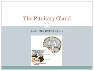
Location • A small gland which lies in the hypophyseal fossa hanging from the hypothalamus, to which it is connected. • Its dimensions are 1 x 1x 1.5 cm and its weight is o.5 gm.
Components • The pituitary consists of two embryologically, histologically and functionally different parts: • Adenohypophysis: from Rathke’s pouch (from oral ectoderm). • Neurohypophysis: from neural ectoderm.
Adenohypophysis • Pars distalis: 75 % of the gland. • Pars intermedia. The two parts are separated by remnant of Rathke’s pouch (epithelial cells which surrounds an amorphous colloid. • Pars tuberalis which surrounds the infundibulum.
Neurohypophysis • Pars nervosa: posterior to pars intermedia. • Infundibulum, connecting pars nervosa to, • Median eminence, which connects the neurohypophysis to the hypothalamus.
Anatomical lobulation • Anterior lobe: • Pars distalis and pars tuberalis. • Posterior lobe: • Pars intermedia and pars nervosa.
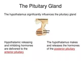
Blood circulation • Superior hypophyseal artery: supplies median eminence and infundibulum (primary capillary plexus) hypophyseal portal circulation pars distalis (secondary capillary plexus). It carries releasing and inhibitory hormones from hypothalamus to adenohypophysis. • Inferior hypophyseal artery: supplies the neurohypophysis. • Hypophyseal veins drain to intracranial venous sinus general circulation.
Function of hypophyseal portal circulation • Hypothalamus produces the neurosecretory hormones which are then stored in the median eminence. Through the hypophyseal portal circulation, these hormones are transferred to the anterior pituitary to affect its cells (either stimulate or inhibit hormone synthesis and secretion). • So, portal circulation is the vascular system used for hormonal regulation of the pars distalis by the hypothalamus.
Hypothalamic neurosecretory hormones (or factors) include: • Thyroid-stimulating hormone releasing hormone (TRH). • Corticotropin-releasing hormone (CRH). • Somatotropin-releasing hormone (SRH). • Gonadotropin-releasing hormone (GnRH). • Prolactin-releasing Hormone (PRH). • Prolactin-inhibitory factor (PIF).
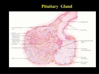
Pars Distalis • Covered by fibrous capsule • Composed of cords of parenchymal cells surrounded by reticular fibrous and sinusoidal capillaries. • Its parenchymal cells are divided into: • Chromophils: • Acidophils. • Basophils. • Chromophobes.
Acidophils • More in number, small rounded cells, having large acidophilic granules. There are two types of acidophils: • Somatotropes: which produce somatotropin (growth hormone) which increases cellular metabolic rate, protein synthesis and elongation of long bones. • Mammotropes: which produce prolactine which promotes mammary gland growth during pregnancy and lactation after delivery.
Basophils • Less in number, located at periphery of pars distalis, There are three types of basophils: • Corticotropes: large, rounded or oval cells with eccentric nuclei, produce adrenocorticotropin (ACTH), lipotropic hormone (LPH). • Thyrotropes: angular in shape, have small granules, produce thyrotropin (TSH). • Gonadotropes: small and rounded in shape, produces: follicular stimulating hormone (FSH), Leuteinizing hormone (LH) and interstitial cell-stimulating hormone (ICSH).
Chromophobes • Small, weakly stained cells. Have less cytoplasm. • The may represent non-specific stem (precursor) cells, or degranulated chromophil cells.
Pars intermedia • Lies between pars distalis and pars nervosa. • Has colloid-containing cysts, which are lined by cuboidal cells and represents remnants of Rathke’s pouch. • Its has basophils arranged in cords around network of capillaries. • These basophils may secrete melanocyte stimulating hormone (MSH).
Pars tuberalis • Surrounds the infundibulum, but it is thinner or may be absent posteriorly. • Formed of longitudinal cords of cuboidal parenchymal basophilic cells surrounded by sinusoids. • These cells have small dense granules and they may share in secretion of FSH and LH.
Neurohypophysis • It develops from downgrowth of the hypothalamus. • It consists of median eminence, infundibulum and pars nervosa. • It is connected to the hypothalamic nuclei (supra-optic and paraventricular nuclei) through the hypothalamo-hypophyseal tract.
It is composed of: • Unmyelinated nerve fibers (forming the hypothalamo-hypophyseal tract). • Herring bodies: are distensions of the axons of the hypothalamo-hypophyseal tracts containing membrane-bound granules full of vasopressin and oxytocin. These granules are released to enter the fenestrated endothelium of the capillaries surrounding them, then to general circulation. • Pituicytes: which are similar to neuroglial cells, they support the axons of the pars nervosa. • Blood capillaries.
Function of pars nervosa • Storing and release of oxytocin which causes contraction of the myoepithelial cells surrounding the alveoli of the mammary glands and contraction of the uterine muscles after labour. • Storage and release of vasopressin (antidiuretic hormone, ADH) which causes reabsorption of water in distal convoluted tubules and collecting tubule of the kidney, and contraction of the smooth muscles of the blood vessels to elevate the blood pressure.