Download
1 / 14
430 likes | 1.69k Vues
Anesthesia Management of Mediastinal Masses. N746 Respiratory Disease Presentation Angela Hickey University of Pennsylvania School of Nursing. Mediastinal Masses. The mediastinum is defined as “the space between the lungs ”

E N D
Anesthesia Management of Mediastinal Masses N746 Respiratory Disease Presentation Angela Hickey University of Pennsylvania School of Nursing
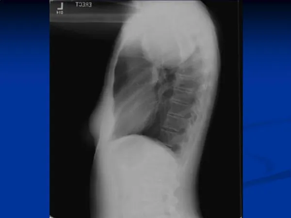
Mediastinal Masses • The mediastinum is defined as “the space between the lungs” • Borders: Thoracic inlet superiorly, Diaphragm inferiorly, Sternum anteriorly, Spine posteriorly, and Pleural spaces laterally. • Divided into compartments: Anterior, Middle, and Posterior • 2/3 of mediastinal masses in adults = anterior • High risk d/t location to the airway and great vessels. • Compress the pulmonary artery and heart • Increase atelectasis and obstruct airflow • CNS changes (d/t obstructed venous drainage in the upper thorax). • GA very dangerous = Collapse of airway= Obstruction • Supine position • Induction of anesthesia • Positive-pressure ventilation
Mediastinal mass • Common AM tumors- ”The 4 T’s”-Thymoma, Thyroid, Teratoma, and “Terrible” Lymphoma • Symptoms of Anterior Mediastinal Mass: • Neuro: syncope • Resp: Stridor, Hoarseness, Cyanosis, Orthopnea, Inability to lie flat, Cough (esp when supine), Chest pain or fullness • CV: Sweating, Jugular Vein Distention, Superior vena cava syndrome • Treatment: depends on type of tumor and symptoms- typically a combination of surgery, chemo, and/or XRT • Surgical procedures can be diagnostic(eg. biopsy for grading), palliative/supportive (eg. stenting of trachea or SVC), or treatment via complete resection of tumor
SVC syndrome • Venous engorgement of the upper body when the SVC is compressed • Typically caused by cancerous tumors • Most common- • Lung and Non-Hodgkin Lymphoma • S/S: dilation of collateral veins of the upper part of the thorax and neck, edema and rubor of the face, neck, upper torso and airway, SOB, HA, visual distortion or altered mentation.
Anesthetic management • Ideally- Local Anesthesia in all circumstances with minimal sedation • If GA necessary: • GOAL= Maintain spontaneous ventilation to retain normal airway-distending pressure gradients and maintain airway patency. • Have rigid bronchoscopy at bedside • Ability to turn patient lateral or prone in case of collapse • Consider cannulation for ECMO if tumor large or symptomatic
Mediastinoscopy • A rigid scope is passed through the suprasternal notch. • Why is it used? For direct visualization of the mediastinal space and to sample paratracheal and mediastinal lymph nodes at various levels in order to stage intrathoracic cancers, lymphoma. • Relatively contraindicated in pts with prior mediastinoscopy, tracheal deviation, radiation, thoracic aneurysm or SVC obstruction d/t increased risk of vessel puncture from anatomic distortion
Mediastinoscopy • Pre-op Considerations • RESP: IMPORTANT evaluations to anticipate potential for airway collapse 1. CT/TEE/MRI- to delineation of mass size and effects 2.Respiratory system assessment: Must be thorough to asses for respiratory tract compression • Symptoms positional? Compare when supine • Asymptomatic or vague signs (dyspnea, cough, hoarseness, or chest pain) • Wheezing (mechanical obstruction vsbronchospasm) • If SVC syndrome: may have significant airway edema • 3. PFTs- helpful-> not always reliable for predicting airway collapse • CV: Vascular compression (Right heart, PA, SVC) Hypotension, hypoxia, SVC syndrome • MS: Assess for myasthenic syndrome (resistance to depolarizing agents and increased sensitivity to NDMRs.) • Neuro: If SVC obstructed, will have increased ICP. Pts with pre-existing carotid disease are at increased risk for stroke
Mediastinoscopy • Pre-op (con’t) • BP monitoring- must occur on left arm (whether invasive or non-invasive) with or without right arm monitoring • Pulse oximeter placed on right arm • 14-16 ga x 1(one in lower extremity with SVC syndrome) • Blood products for transfusion available in OR • CVP with PA line for those with large mediastinal masses (placed via femoral vein with SVC syndrome) • Rigid Bronchoscopy on standby • Difficult airway cart with flexible bronchoscopy on standby • Femoral cannulation for highest-risk patients
MEDIASTINOSCOPY • Induction • Awake fiberoptic using local anesthesia preferred • Allows direct visualization of obstruction t/f ETT placed distal • Monitors the effects of positional changes on the trachea/bronchii • Alternatively, Inhalation induction with Sevo/O2 • ETT should be reinforced if known airway compression • Semi-Fowler’s position • Maintain spontaneous ventilation • Avoid muscle relaxation, if necessary utilize shorter acting only after guaranteed PPV
Mediastinoscopy • Intra-Op Considerations • ***Acute brachiocephalic compression with the scope • Results in hypoperfusion of the R common carotid -> CVA • Innominate artery compression is monitored via pulsatile waveform in the Right upper extremity using pulse oximetry • Left radial arterial line so that blood pressure can be monitored in the event of brachiocephalic avulsion • Most common COMPLICATION*** Hemorrhage d/t laceration of great vessel • Other complications: • Venous Air Embolism- risk higher d/t HOB at 30 degrees and when spontaneously ventilating • Tracheal or Esophageal compression/Injury • Bradycardia (vagally-mediated d/t compression on trachea or great vessels), Arrhythmias, Cardiac Arrest • Thoracic Duct Injury
Mediastinoscopy • Post-op Considerations • Monitor for Complications • Left recurrent laryngeal nerve damage • Phrenic Nerve Injury • Pneumothorax/Pneumopericardium/Pneumomediastinum • Tracheamalacia
Case Study: LMA use in Patient with mediastinal mass • Preop: • 64 yo F with recurrent 10 x10 x12 cm lung cancer tumor encroaching the great vessels and trachea with significant tracheal compression and deviation not amenable to surgery. Scheduled for palliative tracheal stent to relieve the airway obstruction 2/2 worsening dyspnea and cough. • Induction, Maintenance, Post-Op • Large bore IV, right radial A-line • Inhalation induction (70% nitrous/30% O2/Sevothen 100% O2/Sevo) in Semi-recumbent position • Classic LMA #5 placed while spontaneous ventilation maintained • GA with O2/Sevo supplemented with low-dose Propofol (25-50mcg/kg/min) • Tracheal stent was deployed with immediate imporvement of tidal volumes. • LMA successfully removed at the end of the procedure and the patient recovered from the surgery uneventfully in the PACU. • Classic LMA provided better access to the subglottic compression • The large internal diameter and lack of epiglottic elevator bars allow easy passage of the stent loader and bronchoscope without interruption of ventilation • Disadvantages: Airway not secure and always the possibility the tumor could compress the trachea distal to the LMA • IMPT = The authors had a rigid bronch available and the cardiopulmonary bypass team on standby
REferences • Barash, P. G., Cullen, B. F., & Stoelting, R. K. (2009). Clinical anesthesia (6th ed.). Philadelphia: Lippincott Williams & Wilkins. • Blank, R.S. & deSouza D.G. (2011). Anesthetic management of patients with an anterior mediastinal mass: Continuing Professional Development. Can J Anesth, 58: 853-867. • Butterworth, J., Mackey, D., & Wasnick, J. (2013). Morgan & Mikhail’s clinical anesthesiology (5th ed.). New York, NY: McGraw-Hill. • Nagelhout, J. & Plaus, K. (2014). Nurse anesthesia (5th ed.). St. Louis, MO: Elsevier Saunders. • Rajagopalan, S. (2014). Anesthetic management of a large mediastinal mass for tracheal stent placement. Rev Bras Anestesiol, http://dxdoi.org/10.1016/j.bjane.2014.01.009.